. 2013 Jan;11(1):60-7.
doi: 10.3171/2012.9.PEDS12268. Epub 2012 Oct 19.

The effect of alpha-v integrin inhibition on the malignant characteristics of medulloblastoma

Affiliations

The effect of alpha-v integrin inhibition on the malignant characteristics of medulloblastoma

Eric M Thompson et al. J Neurosurg Pediatr. 2013 Jan.

Abstract

Object: Hypoxia induces an aggressive phenotype in some brain tumors in part due to hypoxia-inducible factor-1α (HIF-1α) and integrin expression. The importance of hypoxia in medulloblastoma is unclear and the interaction of HIF-1α and c-Myc in medulloblastoma has not been explored. The objective of this study was to determine if hypoxia induces an aggressive phenotype in human medulloblastoma cells that constitutively express high (D283 Med) or low (DAOY) levels of c-Myc and to determine if blocking α(v) integrins with the monoclonal antibody intetumumab inhibits hypoxia-induced cellular stress responses.

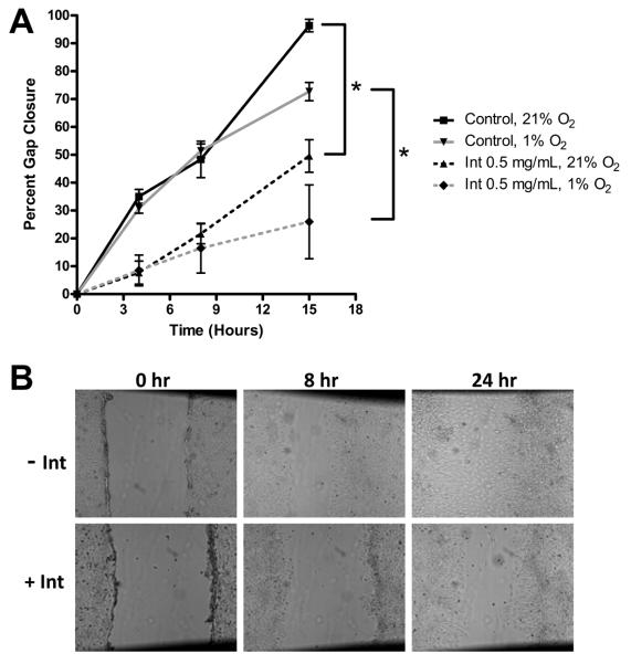
Methods: Cells were grown at 21% and 1% O(2) and in the presence or absence of intetumumab. Measures of malignancy evaluated included cell proliferation, cell migration, and expression of vascular endothelial growth factor (VEGF), α(v) integrins, HIF-1α, and c-Myc.

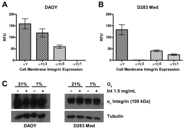
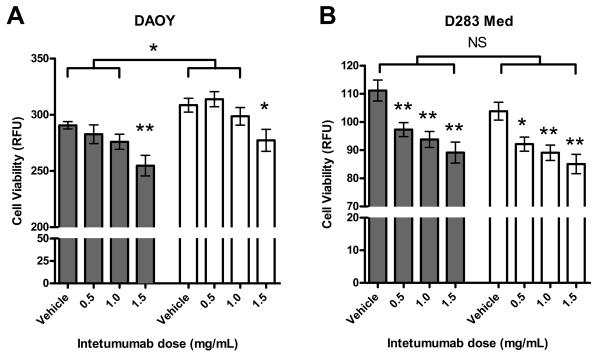
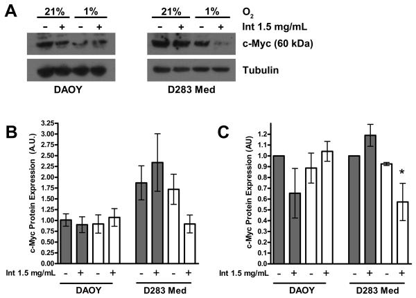
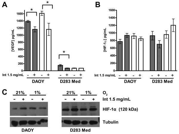
Results: Both cell lines robustly expressed α(v) integrins. Hypoxic DAOY cells showed significantly increased proliferation compared with normoxic controls (p < 0.05), whereas D283 Med cells did not. Both cell lines exhibited a dose-dependent decrease in proliferation when treated with intetumumab (p < 0.05). Hypoxia did not increase DAOY migration, but intetumumab significantly inhibited migration at both oxygen conditions (p < 0.05). Intetumumab significantly decreased VEGF levels in DAOY cells at both oxygen conditions (p < 0.05) and in normoxic D283 cells (p < 0.01). Neither cell line demonstrated increased HIF-1α expression in response to hypoxia. However, hypoxic D283 Med cells grown in the presence of intetumumab demonstrated significantly decreased c-Myc expression (p < 0.05).

Conclusions: Hypoxia did not clearly induce a more aggressive phenotype in medulloblastoma cells. Despite this result, intetumumab decreased medulloblastoma cell proliferation and migration and variably decreased VEGF and c-Myc expression in hypoxic conditions. Targeting α(v) integrins represents a promising potential adjuvant modality in the treatment of medulloblastoma, particularly subtypes that metastasize and overexpress VEGF and c-Myc.


Figures

Figure 1
Figure 1
DAOY and D283 Med cells express αv integrins. A. DAOY cells express αv, αvβ3 and αvβ5 integrins on the cell surface in vitro. B. D283 Med cells express αv, αvβ5 and αvβ1 integrins on the cell surface in vitro. Data represent the mean of 2 different experiments. Values are adjusted to the background level of fluorescence. C. Representative Western blot demonstrates robust expression of αv integrins in both DAOY and D283 Med cell lines. There was no significant difference in the amount of αv integrin expression between the two cell lines, between cells grown at 1% and 21% O2, or cells treated with intetumumab or vehicle. Error bars are standard error. RFU= relative fluorescent unit, Int = intetumumab.
Figure 2
Figure 2
Proliferative response to hypoxia differs in DAOY and D283 Med cells and is inhibited by intetumumab. A. DAOY cells grown in 1% (white bars) or 21% O2 (grey bars) in the presence of 3 escalating doses of intetumumab. Cells grown in the presence of 1.5 mg/mL intetumumab had significantly less proliferation compared to cells grown in the presence of vehicle in their respective oxygen condition. Cells grown at 1% O2 had significantly more proliferation compared to their counterparts grown at 21% O2 with the exception of cells grown in the presence of 1.5 mg/mL intetumumab. B. D283 Med cells grown under 1% or 21% O2 conditions at 3 escalating doses of intetumumab. Cells grown in the presence of all doses of intetumumab had significantly less proliferation compared to cells grown in the presence of vehicle within their respective oxygen condition. There was no difference in proliferation between cells grown at 1% and 21% O2. Figures are means of 3 different experiments. Error bars are standard error. RFU= relative fluorescent unit, * = P < 0.05, ** = P < 0.01, NS = not significant.
Figure 3
Figure 3
c-Myc expression in D283 Med cells is inhibited by the combination of hypoxia and intetumumab. A. Representative Western blot demonstrates decreased expression of c-Myc in DAOY cells compared to D283 Med cells and D283 Med cells grown at 1% in the presence of intetumumab. B. Expression of c-Myc in 3 separate experiments normalized to tubulin appear greater in D283 Med cells than DAOY cells although this difference was not significant due to large variance. C. Expression of c-Myc in 3 separate experiments, normalized to tubulin and to the control (21% O2 vehicle). Neither hypoxic conditions nor the presence of intetumumab significantly affected the expression of c-Myc in DAOY cells. D283 Med cells grown in 1% O2 in the presence of intetumumab had significantly less c-Myc expression compared to controls. Error bars are standard error. Clear bars = 1% O2, grey bars = 21% O2, * = P < 0.05, Int = intetumumab.
Figure 4
Figure 4
Intetumumab inhibits VEGF but not HIF-1α expression in DAOY and D283 Med cells. A. Intetumumab significantly decreased VEGF production in DAOY cells grown at both oxygen conditions and D283 Med cells grown at 21% O2 in 2 separate experiments. B. There was no significant difference in the production of HIF-1α in cells grown in the presence of intetumumab or hypoxic conditions in both DAOY and D283 Med cell lines. C. Representative Western blot demonstrates no significant difference in HIF-1α expression in both cell lines grown in various conditions. Error bars are standard error. Grey bars = 21% O2, clear bars = 1% O2,* = P < 0.05, Int = intetumumab.
Figure 5
Figure 5
Cell migration is inhibited by intetumumab. A. DAOY cell migration in both hypoxic and normoxic conditions was significantly inhibited by intetumumab. There was no difference in rate of migration between the two oxygen environments. B. Representative photos (50 ×) of intetumumab inhibiting cell migration (gap closure) at 0, 8, and 24 hour time points. * = P < 0.05, Int = intetumumab.

Similar articles

Cited by

References

    1. Brandsma D, Ulfman L, Reijneveld JC, Bracke M, Taphoorn MJ, Zwaginga JJ, et al. Constitutive integrin activation on tumor cells contributes to progression of leptomeningeal metastases. Neuro Oncol. 2006;8:127–136. - PMC - PubMed
    1. Brat DJ, Castellano-Sanchez AA, Hunter SB, Pecot M, Cohen C, Hammond EH, et al. Pseudopalisades in glioblastoma are hypoxic, express extracellular matrix proteases, and are formed by an actively migrating cell population. Cancer Res. 2004;64:920–927. - PubMed
    1. Chu FM, Picus J, Fracasso PM, Dreicer R, Lang Z, Foster B. A phase 1, multicenter, open-label study of the safety of two dose levels of a human monoclonal antibody to human alpha(v) integrins, intetumumab, in combination with docetaxel and prednisone in patients with castrate-resistant metastatic prostate cancer. Invest New Drugs. 2011;29:674–679. - PubMed
    1. Fiorilli P, Partridge D, Staniszewska I, Wang JY, Grabacka M, So K, et al. Integrins mediate adhesion of medulloblastoma cells to tenascin and activate pathways associated with survival and proliferation. Lab Invest. 2008;88:1143–1156. - PMC - PubMed
    1. Friedlander M, Brooks PC, Shaffer RW, Kincaid CM, Varner JA, Cheresh DA. Definition of two angiogenic pathways by distinct alpha v integrins. Science. 1995;270:1500–1502. - PubMed

Publication types

MeSH terms

Substances

LinkOut - more resources