

Versican expression in canine carcinomas in benign mixed tumours: is there an association with clinical pathological factors, invasion and overall survival?
- PMID: 23082892
- PMCID: PMC3534148
- DOI: 10.1186/1746-6148-8-195
Versican expression in canine carcinomas in benign mixed tumours: is there an association with clinical pathological factors, invasion and overall survival?
Abstract
Background: Components of the extracellular matrix have been studied in an attempt to elucidate the mechanisms involved in the biological behaviour of tumours. The presence of the proteoglycan versican has been strongly associated with cancer development and progression. However, relationship between versican expression and clinical pathological factors and overall survival has not been previously studied in veterinary medicine. Carcinomas in benign mixed tumours (CBMTs) are one of the most common malignant tumours in female canines and can serve as models for studies of tumour progression. The aim of this study was to evaluate the expression of versican in in situ and invasive carcinomatous areas of canine CBMTs and to evaluate possible associations of versican expression with other classic prognostic factors and overall survival.
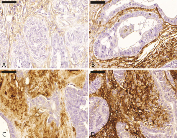
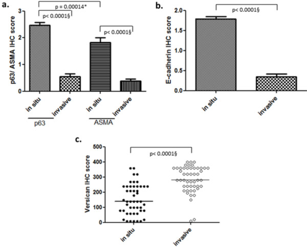
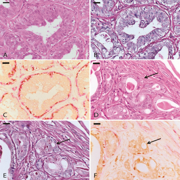
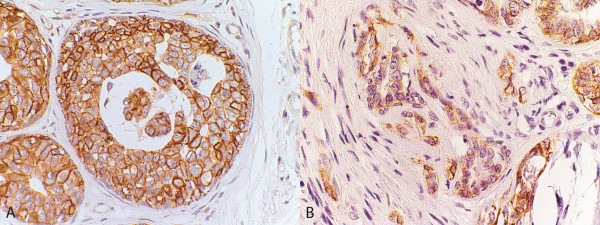
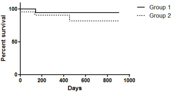
Results: Clinical staging; histological grade determination; immunohistochemical staining for versican, E-cadherin and Ki-67; and confirmation of invasion areas by staining for p63 and smooth muscle α-actin (α-SMA) were performed on 49 canine cases of CBMT. Tumour invasion was considered when suspicious Haematoxylin-Eosin (HE)-stained areas showed a total loss of α-SMA and p63 immunoreactivity. Versican immunoreactivity was less intense in the areas adjacent to the in situ carcinomatous regions, compared to invasive regions, which showed extensive and strong staining.
Conclusions: Our data reveal that in canine CBMTs, versican expression differs significantly between invasive and in situ areas, suggesting a role for this molecule in tumour progression. Although a direct relationship exists between versican and invasiveness, our results indicate that the isolated evaluation of this proteoglycan does not represent an independent prognostic factor in canine CBMTs.
Figures
References
-
- Peleteiro MC. Tumores mamários na cadela e na gata. Rev Port Ciênc Vet. 1994;89: 509:10–29.
-
- Morrison WB. Cancers in dogs and cats, Medical and surgical management. Philadelphia: Willians e Wilkins; 1998. p. 785.
-
- Cassali GD, Melo BM, Madureira N, Ferreira E, Bertagnolli AC, Ribeiro GM, Campos CB. Mammary gland diagnosis of the Laboratory of Comparative Pathology - UFMG, from 2000 to 2008. Clínica Veterinária. 2009;14:173–173.
-
- Estrela-Lima A, Araújo MSS, Costa-Neto JM, Teixeira-Carvalho A, Barrouin-Melo SM, Cardoso SV, Martins-Filho OA, Serakides R, Cassali GD. Immunophenotypic features of tumor infiltrating lymphocytes from mammary carcinomas in female dogs associated with prognostic factors and survival rates. BMC Cancer. 2010;10:1–14. doi: 10.1186/1471-2407-10-1. - DOI - PMC - PubMed
Publication types
MeSH terms
Substances
LinkOut - more resources
Full Text Sources
