. 2013 Mar;168(6):1471-84.
doi: 10.1111/bph.12024.

(E)-2,4-bis(p-hydroxyphenyl)-2-butenal has an antiproliferative effect on NSCLC cells induced by p38 MAPK-mediated suppression of NF-κB and up-regulation of TNFRSF10B (DR5)

Affiliations

(E)-2,4-bis(p-hydroxyphenyl)-2-butenal has an antiproliferative effect on NSCLC cells induced by p38 MAPK-mediated suppression of NF-κB and up-regulation of TNFRSF10B (DR5)

Pushpa Saranya Kollipara et al. Br J Pharmacol. 2013 Mar.

Abstract

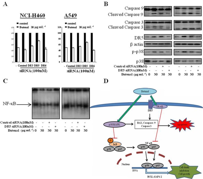
Background and purpose: The Maillard Reaction Products (MRPs) are known to be effective in chemoprevention. Here we focused on the anticancer effects of (E)-2,4-bis(p-hydroxyphenyl)-2-butenal (a MRP) on human non-small-cell lung cancer (NSCLC) cells and its mechanism of action.

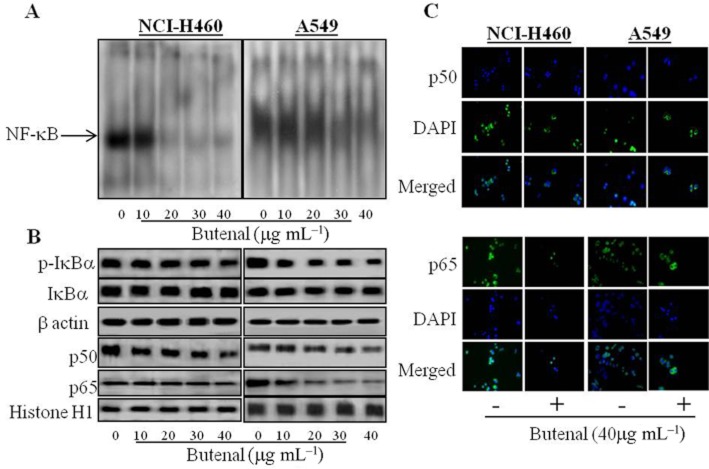
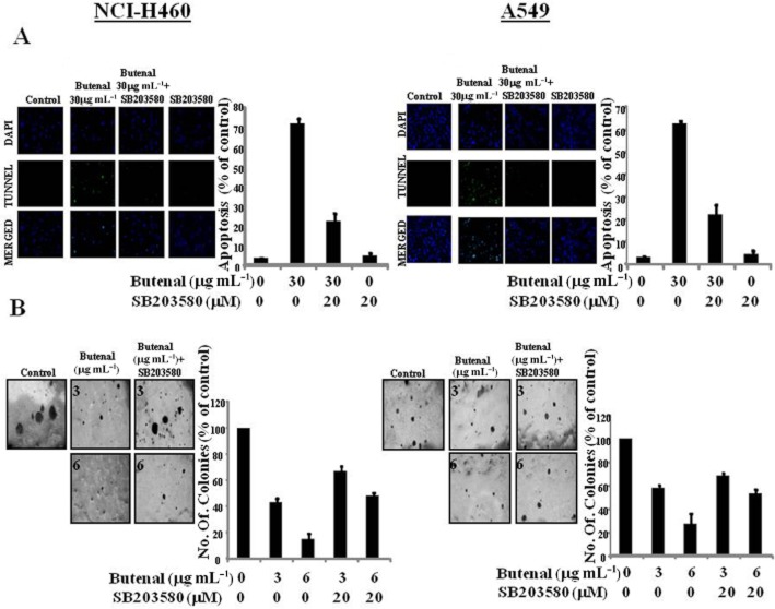
Experimental approach: We analysed the activity of (E)-2,4-bis(p-hydroxyphenyl)-2-butenal on NSCLC cells (NCI-H460 and A549) by use of Western blot analysis for major apoptotic proteins, MAPK, NF-κB and death receptor expression. We also used RT-PCR to determine its effects on death receptor mRNA expression, EMSA for effects on NF-κB DNA binding activity and colony formation assay for effects of inhibitors on (E)-2,4-bis(p-hydroxyphenyl)-2-butenal's actions.

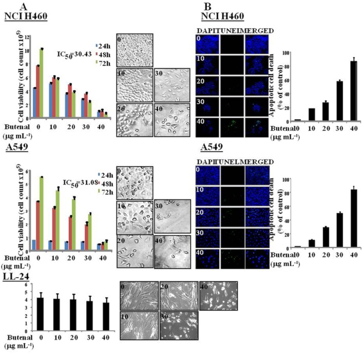
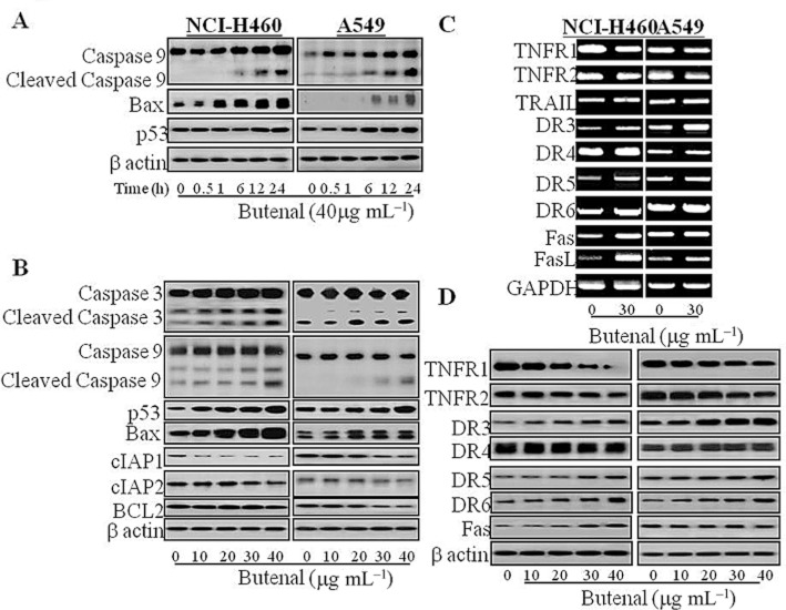
Key results: (E)-2,4-bis(p-hydroxyphenyl)-2-butenal induced a concentration (10-40 μg·mL⁻¹)- and time (30 min-72 h)-dependent inhibitory effect on the growth of NSCLC cells due to induction of apoptosis. Concomitantly, it significantly increased the expression of apoptotic proteins such as cleaved caspase-3, cleaved caspase-9, Bax and p53, but down-regulated the expression of anti-apoptotic proteins Bcl-2, cIAP1 and cIAP2. This effect was induced by up-regulation of MAPK and death receptor proteins TNFRSF12, TNFRSF10B and TNFRSF21, but suppression of NF-κB. Of the death receptors activated, only TNFRSF10B knock down with siRNA reversed the effect of (E)-2,4-bis(p-hydroxyphenyl)-2-butenal. Even though all the MAPKs were activated, only pretreatment with a p38 MAPK inhibitor reversed (E)-2,4-bis(p-hydroxyphenyl)-2-butenal-induced cell growth inhibition, increase in cleaved caspase-3, -9 and TNFRSF10B expression, and NF-κB inactivation.

Conclusions and implications: (E)-2,4-bis(p-hydroxyphenyl)-2-butenal induces apoptosis in NSCLC cells by p38 MAPK-mediated suppression of NF-κB and activation of TNFRSF10B, which then activates the caspase-3 and caspase-9 pathways.


Figures

Figure 1
Figure 1
Effect of (E)-2,4-bis(p-hydroxyphenyl)-2-butenal on the cell viability and morphological changes of NSCLC cell lines. (A) Concentration- and time-dependent effect of (E)-2,4-bis(p-hydroxyphenyl)-2-butenal on cell viability of NCI-H460, A549 and LL-24 cells. After treatment with (E)-2,4-bis(p-hydroxyphenyl)-2-butenal (0–40 μg·mL−1) for 24, 48 and 72 h, the morphological changes of NCI-H460 and A549 were observed and the number of viable cells was counted under a microscope (magnification, ×200). Values are mean ± SD of three experiments with replicates. *P ≤ 0.05 indicates statistically significant differences from the control group. (B) Apoptotic cell death of NSCLC cell lines with the treatment of (E)-2,4-bis(p-hydroxyphenyl)-2-butenal. Quantification of apoptosis by TUNEL assay. The green colour in the fixed cells marks TUNEL-labelled cells. Total number of cells in a given area was determined by using DAPI nuclear staining (fluorescent microscope). The apoptotic index was determined as the DAPI-stained TUNEL-positive cell number/total DAPI-stained cell number (magnification, ×200). Values are mean and SD of three experiments with replicates. *P ≤ 0.05 indicates statistically significant differences from the control group.
Figure 2
Figure 2
Effect of (E)-2,4-bis(p-hydroxyphenyl)-2-butenal on the expression of apoptosis regulatory proteins. (A) The cells were treated with same concentration (40 μg·mL−1) of (E)-2,4-bis(p-hydroxyphenyl)-2-butenal and harvested at different time points. (B) The cells were treated with different concentrations (0–40 μg·mL−1) of (E)-2,4-bis(p-hydroxyphenyl)-2-butenal at 37°C for 12 h. Equal amounts of total proteins (50 μg per lane) were subjected to 10% SDS-PAGE. Expressions of cleaved caspase-3, cleaved caspase-9, p53, Bax, cIAP1/2, Bcl-2 and β-actin were detected by Western blotting using specific antibodies. β-Actin protein was used an internal control in NCI-H460 and A549 lung cancer cells. Each band is representative of three independent experiments. (C) Effect of (E)-2,4-bis(p-hydroxyphenyl)-2-butenal on death receptor expression in NSCLC cell lines. The cells were treated with different concentrations (0–40 μg·mL−1) of (E)-2,4-bis(p-hydroxyphenyl)-2-butenal at 37°C, and total RNA were extracted and examined for expressions of TNFRSF1A and 1B, TNF-R1 and 2, FAS, TNFRSF12, 10A, 10B and 21 (DR-3, -4, -5, -6) and GAPDH by RT-PCR. GAPDH was used as an internal control to show equal RNA loading. Equal amounts of total proteins (50 μg·per lane) were subjected to 10% SDS-PAGE. Expressions of TNFRSFs, FAS, and β-actin were detected by Western blotting using specific antibodies. β-Actin protein was used an internal control. Each band is representative for three experiments.
Figure 3
Figure 3
Effect of (E)-2,4-bis(p-hydroxyphenyl)-2-butenal on NF-κB activation in NSCLC cell lines. (A) Nuclear extracts from NCI-H460 and A549 lung cancer cells treated with (E)-2,4-bis(p-hydroxyphenyl)-2-butenal (0–40 μg·mL−1) for 1 h were incubated in binding reactions of 32P-end-labelled oligonucleotide containing the κB sequence. The activation of NF-κB was investigated using EMSA as described in Methods. (B) The cells treated with (E)-2,4-bis(p-hydroxyphenyl)-2-butenal (0–40 μg·mL−1) for 1 h were lysed, cytosolic proteins were used to determine the expression of IκB, p-IκB and β-actin (internal control) and nuclear proteins were used to determine the expression of p50, p65 and histoneH1(internal control) in lung cancer cells. (C) Intracellular location of p50 and p65 in lung cancer cells was determined by immunofluorescence confocal scanning microscope (magnification, 400×). Double staining (Merge) with p50 or p65 and DAPI staining demonstrates the localization of p50 and p65 in the nucleus. Values are mean ± SD of three experiments with replicates. *P ≤ 0.05 indicates statistically significant differences from the control group.
Figure 4
Figure 4
Effect of (E)-2,4-bis(p-hydroxyphenyl)-2-butenal on MAPK activation in NSCLC cell lines. (A) The cells were treated with different concentrations (0–40 μg·mL−1) of (E)-2,4-bis(p-hydroxyphenyl)-2-butenal at 37°C of 30 min, equal amounts of total proteins (50 μg per lane) were subjected to 10% SDS-PAGE. Expressions of ERK, p-ERK, JNK, p-JNK, p38, p-p38 and β-actin were detected by Western blotting using specific antibodies. β-Actin protein was used an internal control in both NCI-H460 and A549 lung cancer cells. Each band is representative of three independent experiments. Effect of p38 inhibitor SB203580 on the activity of (E)-2,4-bis(p-hydroxyphenyl)-2-butenal. (B) The effect of pretreatment with SB203580 on the (E)-2,4-bis(p-hydroxyphenyl)-2-butenal-induced decrease in cell viability. After treatment with (E)-2,4-bis(p-hydroxyphenyl)-2-butenal (0–40 μg·mL−1) for 24 h, the cells were harvested by trypsinization and stained with 0.2% trypan blue. Relative cell survival rate was determined by counting live and dead cells. (C) The effect of pretreatment with SB203580 on the (E)-2,4-bis(p-hydroxyphenyl)-2-butenal-induced increase in the death receptor expression and caspase-3 and -9 activation. (D) The effect of pretreatment with SB203580 on the (E)-2,4-bis(p-hydroxyphenyl)-2-butenal-induced NF-κB inactivation, the cells treated with (E)-2,4-bis(p-hydroxyphenyl)-2-butenal (0–40 μg·mL−1) for 1 h were lysed, cytosolic proteins were used to determine the expression of IκB, p-IκB and β-actin (internal control). The effect of pretreatment with SB203580 on the (E)-2,4-bis(p-hydroxyphenyl)-2-butenal-induced inhibition of NF-κB DNA binding activity. Nuclear extracts from NCI-H460 and A549 lung cancer cells pretreated with SB203580 for 30 min and treated with (E)-2,4-bis(p-hydroxyphenyl)-2-butenal (0–40 μg·mL−1) for 1 h were incubated in binding reactions of 32P-end-labelled oligonucleotide containing the κB sequence. The activation of NF-κB was investigated using EMSA as described in Methods. Values are mean ± SD of three experiments with replicates. *P ≤ 0.05 indicates statistically significant differences from the control group.
Figure 5
Figure 5
(A) The effect of pretreatment with SB203580 on the (E)-2,4-bis(p-hydroxyphenyl)-2-butenal-induced apoptosis, the apoptotic cell death of non-small cell lung cancer cell lines pretreated with SB203580 and then treated with (E)-2,4-bis(p-hydroxyphenyl)-2-butenal. Quantification of apoptosis by TUNEL assay. The NSCLC cells NCI-H460 and A549, were pretreated with 20 μM SB203580 for 30 min and then treated with (E)-2,4-bis(p-hydroxyphenyl)-2-butenal (30 μg·mL−1) for 24 h and then labelled with TUNEL solution. The green colour in the fixed cells marks TUNEL-labelled cells. Total number of cells in a given area was determined by using DAPI nuclear staining (fluorescent microscope). The apoptotic index was determined as the DAPI-stained TUNEL-positive cell number/total DAPI-stained cell number (magnification, ×200). Values are mean ± SD of three experiments with replicates. *P ≤ 0.05 indicates statistically significant differences from the control group. (B) The effect of pretreatment with SB203580 on the (E)-2,4-bis(p-hydroxyphenyl)-2-butenal-induced inhibition of colony formation in NCI-H460 and A549 cells. After 30 min of pretreatment 8 × 10³ cell·mL−1 were suspended in 2 mL of 0.3% agar containing basal medium Eagle's agar containing 10% FBS. The cultures were maintained at 37°C in a 5% CO2 atmosphere for 2 weeks, and cell colonies >80 μm in diameter were scored. Values are mean ± SD of three experiments with replicates. *P ≤ 0.05 indicates statistically significant differences from the control group.
Figure 6
Figure 6
Effect of siRNA of TNFRSFs (death receptors) on the (E)-2,4-bis(p-hydroxyphenyl)-2-butenal-induced cancer cell growth inhibition, p38 expression and apoptosis in lung cancer cells. (A) Effect of siRNA of DR on the (E)-2,4-bis(p-hydroxyphenyl)-2-butenal-induced cell viability. The lung cancer cells were transfected with the DR siRNA (100 nM) for 24 h; the cells were then treated with (E)-2,4-bis(p-hydroxyphenyl)-2-butenal (30 μg·mL−1) for another 24 h. The cells were harvested by trypsinization and stained with 0.2% trypan blue. Relative cell survival rate was determined by counting live and dead cells. Values are mean ± SD of three experiments with replicates. *P ≤ 0.05, significantly different from untreated control cells. #P ≤ 0.01, significantly different from control siRNA transfected cells. (B) The cells were transfected with non-targeting control siRNA or TNFRSF10B (DR5) siRNA (100 nM) as described in Methods for 24 h. Then (E)-2,4-bis(p-hydroxyphenyl)-2-butenal was treated (30 μg·mL−1) at 37°C. Equal amounts of total proteins (50 μg·per lane) were subjected to 10% SDS-PAGE. Expressions of cleaved caspase-3, cleaved caspase-9, TNFRSF10B, p38, p-p38 and β-actin were detected by Western blotting using specific antibodies. β-Actin protein was used an internal control in NCI-H460 and A549 lung cancer cells. Each band is representative of three independent experiments. (C) Effect of siRNA of TNFRSF10B (DR5) on the (E)-2,4-bis(p-hydroxyphenyl)-2-butenal-induced NF-κB inactivation. Nuclear extract from NCI-H460 and A549 lung cancer cells pre-treated with SB203580 of 30 min and treated with (E)-2,4-bis(p-hydroxyphenyl)-2-butenal (0–40 μg·mL−1) for 1 h was incubated in binding reactions of 32P-end-labelled oligonucleotide containing the κB sequence. The activation of NF-κB was investigated using EMSA as described in Methods. Values are mean ± SD of three experiments with replicates. *P ≤ 0.05 indicates statistically significant differences from the control group. (D) Schematic representation of anti-proliferative effect of (E)-2,4-bis(p-hydroxyphenyl)-2-butenal on NSCLC cells.

Similar articles

Cited by

References

    1. Ashkenazi A. Targeting death and decoy receptors of the tumor-necrosis factor super family. Nat Rev Cancer. 2002;2:420–430. - PubMed
    1. Atrooz OM. The effects of Maillard reaction products on apple and potato polyphenoloxidase and their antioxidant activity. Int J Food Sci Technol. 2008;43:490–494.
    1. Baud V, Karin M. Signal transduction by tumor necrosis factor and its relatives. Trends Cell Biol. 2001;11:372–377. - PubMed
    1. Baud V, Karin M. Is NF-kappaB a good target for cancer therapy? Hopes and pitfalls. Nat Rev Drug Discov. 2009;8:33–40. - PMC - PubMed
    1. Bian J, Wang K, Kong X, Liu H, Chen F, Hu M, et al. Caspase- and p38-MAPK-dependent induction of apoptosis in A549 lung cancer cells by Newcastle disease virus. Arch Virol. 2011;156:1335–1344. - PubMed

Publication types

MeSH terms