

Effects of myocardial infarction on the distribution and transport of nutrients and oxygen in porcine myocardium
- PMID: 23083196
- PMCID: PMC3625428
- DOI: 10.1115/1.4007455
Effects of myocardial infarction on the distribution and transport of nutrients and oxygen in porcine myocardium
Abstract
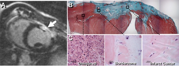
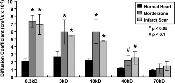
One of the primary limitations of cell therapy for myocardial infarction is the low survival of transplanted cells, with a loss of up to 80% of cells within 3 days of delivery. The aims of this study were to investigate the distribution of nutrients and oxygen in infarcted myocardium and to quantify how macromolecular transport properties might affect cell survival. Transmural myocardial infarction was created by controlled cryoablation in pigs. At 30 days post-infarction, oxygen and metabolite levels were measured in the peripheral skeletal muscle, normal myocardium, the infarct border zone, and the infarct interior. The diffusion coefficients of fluorescein or FITC-labeled dextran (0.3-70 kD) were measured in these tissues using fluorescence recovery after photobleaching. The vascular density was measured via endogenous alkaline phosphatase staining. To examine the influence of these infarct conditions on cells therapeutically used in vivo, skeletal myoblast survival and differentiation were studied in vitro under the oxygen and glucose concentrations measured in the infarct tissue. Glucose and oxygen concentrations, along with vascular density were significantly reduced in infarct when compared to the uninjured myocardium and infarct border zone, although the degree of decrease differed. The diffusivity of molecules smaller than 40 kD was significantly higher in infarct center and border zone as compared to uninjured heart. Skeletal myoblast differentiation and survival were decreased stepwise from control to hypoxia, starvation, and ischemia conditions. Although oxygen, glucose, and vascular density were significantly reduced in infarcted myocardium, the rate of macromolecular diffusion was significantly increased, suggesting that diffusive transport may not be inhibited in infarct tissue, and thus the supply of nutrients to transplanted cells may be possible. in vitro studies mimicking infarct conditions suggest that increasing nutrients available to transplanted cells may significantly increase their ability to survive in infarct.
Figures


References
-
- American Heart Association (AHA), 2006, “Heart Disease and Stroke Statistics—2006 Update,” http://circ.ahajournals.org/cgi/content/short/113/116/e185 - PubMed
-
- Menasche, P. , 2003, “Myoblast-Based Cell Transplantation,” Heart Fail. Rev. 8, pp. 221–227. - PubMed
-
- Schachinger, V. , Assmus, B. , Honold, J. , 2006, “Normalization of Coronary Blood Flow in the Infarct-Related Artery After Intracoronary Progenitor Cell Therapy: Intracoronary Doppler Substudy of the TOPCARE-AMI Trial,” Clin. Res. Cardiol., 95, pp. 13–22. - PubMed
Publication types
MeSH terms
Substances
Grants and funding
LinkOut - more resources
Full Text Sources
Medical
