

The physical foundation of vasoocclusion in sickle cell disease
- PMID: 23083726
- PMCID: PMC3475338
- DOI: 10.1016/j.bpj.2012.09.003
The physical foundation of vasoocclusion in sickle cell disease
Abstract
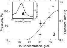
The pathology of sickle cell disease arises from the occlusion of small blood vessels because of polymerization of the sickle hemoglobin within the red cells. We present measurements using a microfluidic method we have developed to determine the pressure required to eject individual red cells from a capillary-sized channel after the cell has sickled. We find that the maximum pressure is only ∼100 Pa, much smaller than typically found in the microcirculation. This explains why experiments using animal models have not observed occlusion beginning in capillaries. The magnitude of the pressure and its dependence on intracellular concentration are both well described as consequences of sickle hemoglobin polymerization acting as a Brownian ratchet. Given the recently determined stiffness of sickle hemoglobin gels, the observed obstruction seen in sickle cell disease as mediated by adherent cells can now be rationalized, and surprisingly suggests a window of maximum vulnerability during circulation of sickle cells.
Copyright © 2012 Biophysical Society. Published by Elsevier Inc. All rights reserved.
Figures

References
-
- Eaton W.A., Hofrichter J., Ross P.D. Editorial. Delay time of gelation: a possible determinant of clinical severity in sickle cell disease. Blood. 1976;47:621–627. - PubMed
-
- Eaton W.A., Hofrichter J. Hemoglobin S gelation and sickle cell disease. Blood. 1987;70:1245–1266. - PubMed
-
- Frenette P.S. Sickle cell vasoocclusion: heterotypic, multicellular aggregations driven by leukocyte adhesion. Microcirculation. 2004;11:167–177. - PubMed
Publication types
MeSH terms
Substances
LinkOut - more resources
Full Text Sources
Medical
