. 2012 Dec;188(6):2063-71.
doi: 10.1016/j.juro.2012.08.030. Epub 2012 Oct 18.

Succinate dehydrogenase kidney cancer: an aggressive example of the Warburg effect in cancer

Affiliations

Succinate dehydrogenase kidney cancer: an aggressive example of the Warburg effect in cancer

Christopher J Ricketts et al. J Urol. 2012 Dec.

Abstract

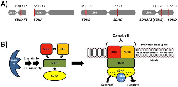
Purpose: Recently, a new renal cell cancer syndrome has been linked to germline mutation of multiple subunits (SDHB/C/D) of the Krebs cycle enzyme, succinate dehydrogenase. We report our experience with the diagnosis, evaluation and treatment of this novel form of hereditary kidney cancer.

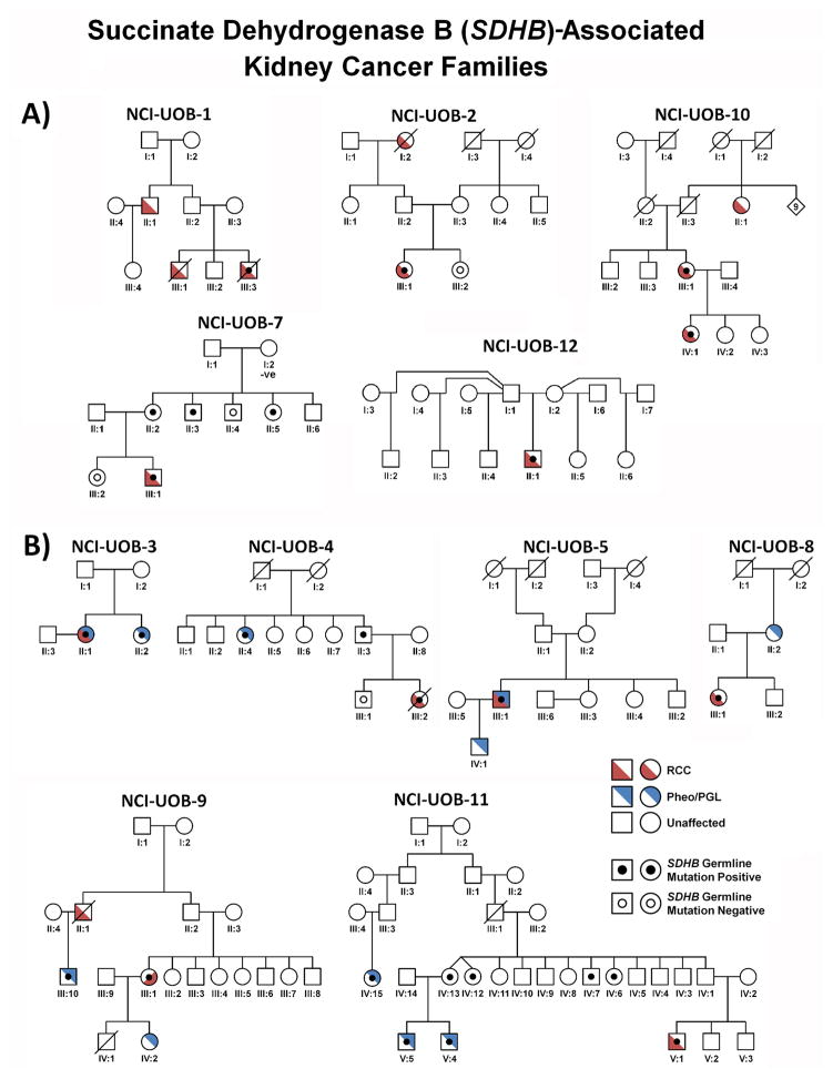
Materials and methods: Patients with suspected hereditary kidney cancer were enrolled on a National Cancer Institute institutional review board approved protocol to study inherited forms of kidney cancer. Individuals from families with germline SDHB, SDHC and SDHD mutations, and kidney cancer underwent comprehensive clinical and genetic evaluation.

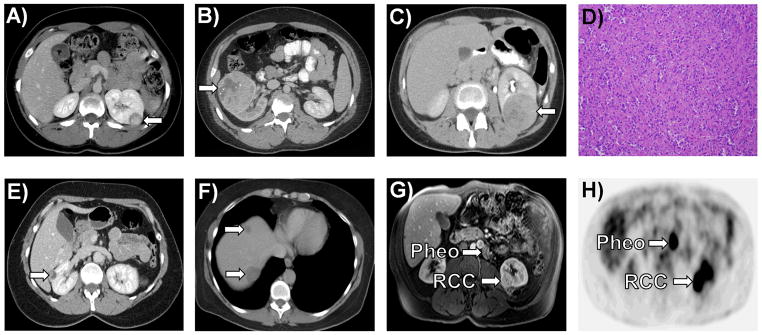
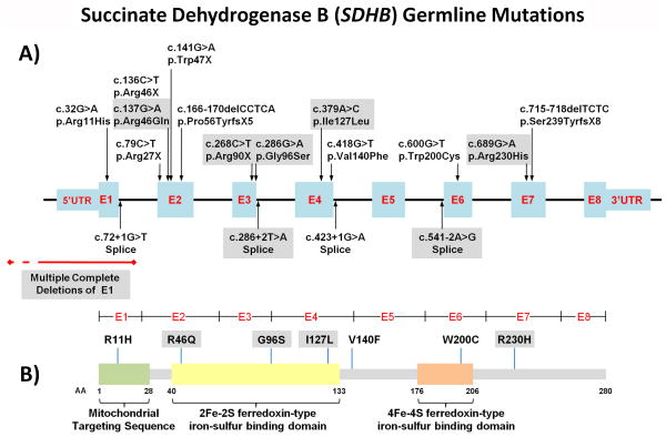
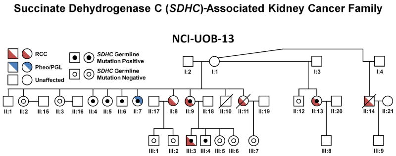
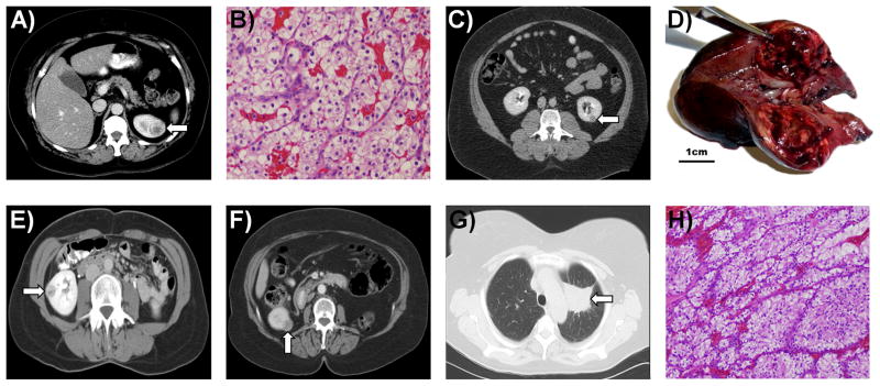
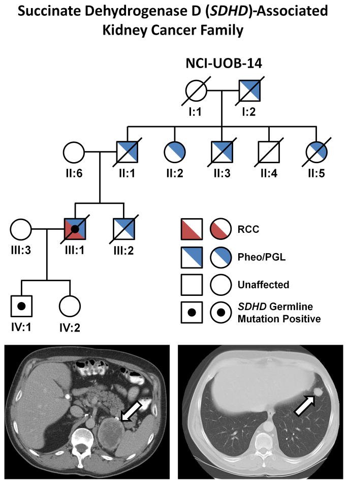
Results: A total of 14 patients from 12 SDHB mutation families were evaluated. Patients presented with renal cell cancer at an early age (33 years, range 15 to 62), metastatic kidney cancer developed in 4 and some families had no manifestation other than kidney tumors. An additional family with 6 individuals found to have clear cell renal cell cancer that presented at a young average age (47 years, range 40 to 53) was identified with a germline SDHC mutation (R133X) Metastatic disease developed in 2 of these family members. A patient with a history of carotid body paragangliomas and an aggressive form of kidney cancer was evaluated from a family with a germline SDHD mutation.

Conclusions: SDH mutation associated renal cell carcinoma can be an aggressive type of kidney cancer, especially in younger individuals. Although detection and management of early tumors is most often associated with a good outcome, based on our initial experience with these patients and our long-term experience with hereditary leiomyomatosis and renal cell carcinoma, we recommend careful surveillance of patients at risk for SDH mutation associated renal cell carcinoma and wide surgical excision of renal tumors.


Figures

Figure 1
Figure 1. The Chromosomal Location of the Succinate Dehydrogenase Enzyme Genes and the Enzyme Assembly
A) Chromosomal mapping of SDHAF1 (19q13.12), SDHA (5p15.33), SDHB (1p36.13), SDHC (1q23.3), SDHAF2 (11q12.2), SDHD (11q23.1). B) The succinate dehydrogenase enzyme subunits (SDHA/B/C/D) are assembled with the aid of the SDHAF1 protein and localized with the mitochondrial membrane, in association with the SDHAF2 protein, to form both Complex II of the Electron Transport Chain and a critical component of the Kreb’s Cycle. (adapted from http://en.wikipedia.org/wiki/File:SDHBfunction.svg)
Figure 2
Figure 2. SDHB-Associated SDH-RCC Family Pedigrees
Pedigrees for SDHB-associated SDH-RCC families (except the adopted patient UOB-6), who was adopted) are shown in two groups: A) families presenting with kidney cancer only and B) families with RCC and pheochromocytoma/paraganglioma. Red denotes RCC and blue denotes pheochromocytoma/paraganglioma (PCC/PGL). SHDB germline mutation positive patients are designated by a black dot and assessed SDHB germline mutation negative patients are designated by an empty black circle.
Figure 3
Figure 3. Imaging and Histology of SDHB-Associated Kidney Cancer
A) A left sided 2.2 cm T1a “oncocytic renal cancer” was detected incidentally in a 19 year old female (UOB-8 III:1). B) A 17 year old male (UOB-12 II:1), who presented with back pain and gross hematuria, underwent a right radical nephrectomy and was found to have a 7.2 cm (T2a) right “oncocytic renal cancer” with sarcomatoid features. C) A 32 year old female (UOB-4 III:2) presented with a 3 month history of back pain and a bone marrow biopsy that revealed ‘metastatic cancer consistent with renal primary”. She underwent a radical nephrectomy for this T3aN0M1 kidney cancer that was diagnosed as an “oncocytic cancer” (D). She died with widespread disease 7 months after surgery. E) Patient UOB-10 III:1 presented as a 55 year old female with a 2.8 cm T1aN0M0 renal mass, which was resected at an outside hospital with clean surgical margins. F) Two and a half years later she was found to have hepatic metastases. G) Patient UOB-5 III:1 came to NCI for routine screening for his family history of SDHB mutation associated disease. On screening he was incidentally found to have a left lower pole 2.8 cm T1aN0M0 renal mass and a 1 cm periaortic paraganglioma. H) Preoperative [18]fluoro-deoxyglucose PET scan was positive for both the renal mass and the paraganglioma. A left partial nephrectomy and excision of the periaortic paraganglioma was performed.
Figure 4
Figure 4. Mutation Map of SDHB mutations associated with SDH-RCC Families
A) Map of SDHB gene mutation in SDHB-associated families with RCC. Exons are shown in blue and introns shown in black lines. SDHB mutations in NCI-UOB families are demonstrated in grey boxes. Previously published mutations are unshaded. Deletions in exon 1 are shown in red. B) SDHB protein mutation map demonstrates missense mutations located throughout the protein with amino acid changes in the mitochondrial targeting sequence as well as both iron-sulfur binding domains.
Figure 5
Figure 5. SDHC-Associated SDH-RCC
The pedigree for the SDHC-associated SDH-RCC family NCI-UOB-13 is shown with the occurrence of RCC (red) and/or pheochromocytoma/paraganglioma (Pheo/PGL) (blue). SHDC germline mutation positive patients are designated by a black dot and assessed SDHC germline mutation negative patients are designated by an empty black circle.
Figure 6
Figure 6. SDHC-Associated Kidney Cancer
A) Patient UOB-13 II:13 was a 49 year old female who presented for screening with a germline SDHC mutation and with 3 sisters affected with kidney cancer. On screening she was found to have a 3.3 cm enhancing left renal lesion. She underwent a partial nephrectomy with a wide surgical margin for removal of a T1aN0M0 clear cell kidney cancer (B). C) Patient UOB-13 III:3 was a 38 year old male who was found to have a germline SDHC mutation. On screening evaluation a 1.4 cm enhancing, exophytic left renal mass was identified. He underwent a retroperitoneal laparoscopic partial nephrectomy with wide surgical margins for a clear cell kidney cancer (D). E) Patient UOB-13 II:8 was a 44 year old female who presented to the NCI for an evaluation of familial renal cancer. She was found to have a 2 cm right renal mass which was removed by right laparoscopic partial nephrectomy and found to be a T1aN0M0 clear cell kidney cancer. She remains free of disease 8 years after surgery. Patient UOB-13 II:9 presented at age 53 with a 10 cm renal tumor (clear cell), for which she had had a left radical nephrectomy. She subsequently underwent a cholecystectomy for a gallbladder recurrence and was found to have a 2.5 cm right renal tumor (F), for which she underwent a partial nephrectomy for a clear cell kidney cancer (H). She was subsequently found to have pulmonary metastases (G) for which she has received systemic therapy.
Figure 7
Figure 7. SDHD-Associated SDH-RCC
A) The pedigree for the SDHD-associated SDH-RCC family (NCI-UOB-14) is shown with the occurrence of RCC (red) and/or pheochromocytoma/paraganglioma (PCC/PGL) (blue). The mutation positive patients are designated by a black dot. Patient III:1 had multiple carotid body tumors and retroperitoneal, extra-adrenal pheochromocytomas (PGL). At age 45 he was found to have an 8 cm left renal mass, which was removed surgically and found to be a T3aNxM1 clear cell renal carcinoma. He was subsequently found to have retroperitoneal (B) and pulmonary (C) recurrence and he died 18 months after the diagnosis of kidney cancer.

Similar articles

Cited by

References

    1. Linehan WM, Srinivasan R, Schmidt LS. The genetic basis of kidney cancer: a metabolic disease. Nature Reviews Urology. 2010;7:277. - PMC - PubMed
    1. Tomlinson IP, Alam NA, Rowan AJ, et al. Germline mutations in FH predispose to dominantly inherited uterine fibroids, skin leiomyomata and papillary renal cell cancer. Nat Genet. 2002;30:406. - PubMed
    1. Grubb RL, III, Franks ME, Toro J, et al. Hereditary leiomyomatosis and renal cell cancer: a syndrome associated with an aggressive form of inherited renal cancer. J Urol. 2007;177:2074. - PubMed
    1. Toro JR, Nickerson ML, Wei MH, et al. Mutations in the fumarate hydratase gene cause hereditary leiomyomatosis and renal cell cancer in families in North America. Am J Hum Genet. 2003;73:95. - PMC - PubMed
    1. Warburg O, Posener K, Negelein E. Metabolism of the Carcinoma Cell. Biochem Z. 1924;152:309.

Publication types

MeSH terms

Substances