Review
. 2012 Dec 20;434(2):187-201.
doi: 10.1016/j.virol.2012.09.026. Epub 2012 Oct 22.

On the nature of mycobacteriophage diversity and host preference

Affiliations
Review

On the nature of mycobacteriophage diversity and host preference

Deborah Jacobs-Sera et al. Virology. .

Abstract

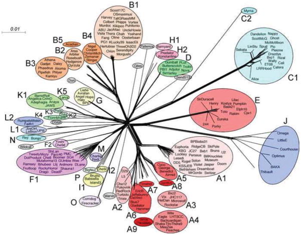
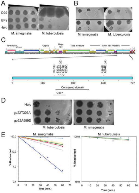
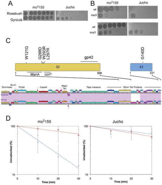
The complete genome sequences of over 220 mycobacteriophages reveal them to be highly diverse, with numerous types sharing little or no nucleotide sequence identity with each other. We have determined the preferences of these phages for Mycobacterium tuberculosis and for other strains of Mycobacterium smegmatis, and find there is a correlation between genome type (cluster, subcluster, singleton) and host range. For many of the phages, expansion of host range occurs at relatively high frequencies, and we describe several examples in which host constraints occur at early stages of infection (adsorption or DNA injection), and phages have the ability to expand their host range through mutations in tail genes. We present a model in which phage diversity is a function of both the ability of phages to rapidly adapt to new hosts and the richness of the diversity of the bacterial population from which those phages are isolated.


Figures

Figure 1
Figure 1
Gene content relationships among mycobacteriophages. The relationships among 220 mycobacteriophages are displayed using the NeighborNet function in Splitstree4 (Huson, 1998). Data were generated from the database Mycobacteriophages_220 in the program Phamerator (Cresawn et al., 2011). Phages within a cluster or subcluster are circled and labeled accordingly. Singletons are shown in grey circles.
Figure 2
Figure 2
Gene content relationships among the subclusters of Cluster A mycobacteriophages. An expanded representation of the Cluster A phages from Figure 1 is shown with each of the subclusters circled and the GC% range for each subcluster indicated.
Figure 3
Figure 3
Host Range Expansion of Cluster G mycobacteriophages. (A) Mycobacteriophages BPs and Halo form plaques on M. tuberculosis mc27000 at an efficiency of 10−4 – 10−5, relative to M. smegmatis mc2155. A control phage, D29, infects both bacteria with equal efficiency. (B) A single Halo plaque picked from an M. tuberculosis and re-plated on M. tuberculosis and M. smegmatis infects both bacteria with equal efficiency (top part). A single plaque picked from the M. smegmatis plate in the top part of the figure retains the ability to efficiently infect M. tuberculosis (bottom part), showing it has acquired a heritable mutation conferring host range expansion. (C) The left arms of the BPs and Halo genomes contain the virion structure and assembly genes (126). The positions of a conserved domain and a weakly predicted coiled domain are shown. (D) Two of the mutations conferring host range expansion – T303A and A599D – were engineered back into mycobacteriophage Halo using BRED mutagenesis. Both mutant phage derivatives efficiently infect M. tuberculosis. (E) Halo and BPs host range mutants [A604E (blue squares); R331Q (purples crosses); T303A (brown squares); A306V (yellow triangles); A321E (green crosses); A599D (red circles) display increased adsorption rates on M. smegmatis relative to wild-type phage (light blue diamonds); adsorption to M. tuberculosis is unaffected.
Figure 4
Figure 4
Isolation and characterization of Rosebush expanded host range mutants. (A) Plating of serial dilutions Rosebush on M. smegmatis mc2155 and M. smegmatis Jucho shows the reduced efficiency of plating; the control phage Qyrzula (which, like Rosebush, is in Subcluster B2). (B) A Rosebush plaque picked from the Jucho plate (mut1) plates on mc2155 and Jucho with equivalent efficiencies of plating (upper). A mut1 plaque recovered from M. smegmatis mc2155 plates with equivalent efficiencies on both strains, in contrast to wild-type Rosebush (wt; lower). (C) Locations of amino acid substitutions in gp32 and gp42 of Rosebush in the expanded host range mutants. Positions of the ManA domain and a weakly predicted coiled domain are shown, as well as a region at the C-terminus of gp32 that is homologous with gp42. (D) Adsorption assays (n = 2) of wild-type Rosebush (red squares) and expanded host range mutant 1 (blue diamonds) on M. smegmatis mc2155 and M. smegmatis Jucho as indicated.
Figure 5
Figure 5
Transfection and replication in non-permissive hosts. (A) Transfection of electrocompetent M. smegmatis Jucho (top row) and mc2155 (bottom row) with 150 ng of either Rosebush genomic DNA (left column) or Giles genomic DNA (right column) followed by recovery and plating with M. smegmatis mc2155 cells. (B) Transfection of electrocompetent M. tuberculosis mc27000 with 150 ng of either Giles genomic DNA (left plate) or D29 genomic DNA (right plate) followed by recovery and plating with M. smegmatis mc2155 cells. (C) Transfection of electrocompetent M. smegmatis mc2155 with either 200 ng (left plate) or 400 ng (right plate) of Streptomyces phage Zemlya followed by recovery and plating with S. lividans. Individual plaques were picked and tested for infection of S. lividans and M. smegmatis; all infected S. lividans but failed to infect M. smegmatis, showing that these are not mutants or contaminants.
Figure 6
Figure 6
A model accounting for mycobacteriophage diversity. The large number of different types of mycobacteriophages isolated on M. smegmatis mc2155 can be explained by a model in which a) phages can readily infect a new bacterial host – either by a switch or an expansion of host range – and b) a highly diverse bacterial population, including many closely related strains, in the environments from which the phages are isolated. As such, phages with distinctly different genome sequences and GC% contents infecting distantly related bacterial hosts – such as those to the left (red) or right (blue) extremes of a spectrum of hosts – can migrate across a microbial landscape through multiple steps. Each host switch occurs at a relatively high frequency (~1 in 105 particles, or an average of about one every 103 bursts of lytic growth), and much faster than either amelioration of phage GC% to its new host, or genetic recombination. Two phages (such as those shown in red and blue) can thus ‘arrive’ at a common host (M. smegmatis mc2155) but be of distinctly different types (clusters, subclusters, and singletons). The variety of hosts is shown two dimensionally for simplicity, and the actual relationships among bacteria in environments such as soil and compost is likely to be considerably more complicated. Because host range switching or expansion is a common feature of bacteriophages, the model predicts that a high degree of phage diversity will be seen for any particular host if the microbial population from which the phages are isolated from is also highly diverse and rich in closely related strains. Because none of the phages isolated on M. smegmatis mc2155 also infect M. aichiense, we assume that this strain and its close relatives are absent from the soil and compost environments where most of these phages were isolated from [M. aichiense was isolated from soil in Japan (Ichiyama et al., 1988)].

References

    1. Abrescia NG, Bamford DH, Grimes JM, Stuart DI. Structure unifies the viral universe. Annu Rev Biochem. 2012;81:795–822. - PubMed
    1. Adriaenssens EM, Mattheus W, Cornelissen A, Shaburova O, Krylov VN, Kropinski AM, Lavigne R. Complete genome sequence of the giant Pseudomonas phage Lu11. J Virol. 2012;86:6369–6370. - PMC - PubMed
    1. Amitsur M, Levitz R, Kaufmann G. Bacteriophage T4 anticodon nuclease, polynucleotide kinase and RNA ligase reprocess the host lysine tRNA. EMBO J. 1987;6:2499–2503. - PMC - PubMed
    1. Arisaka F, Kanamaru S, Leiman P, Rossmann MG. The tail lysozyme complex of bacteriophage T4. Int J Biochem Cell Biol. 2003;35:16–21. - PubMed
    1. Bae B, Ohene-Adjei S, Kocherginskaya S, Mackie RI, Spies MA, Cann IK, Nair SK. Molecular basis for the selectivity and specificity of ligand recognition by the family 16 carbohydrate-binding modules from Thermoanaerobacterium polysaccharolyticum ManA. J Biol Chem. 2008;283:12415–12425. - PubMed

Publication types