. 2013 Jan 20;435(2):463-71.
doi: 10.1016/j.virol.2012.09.042. Epub 2012 Oct 16.

Contribution of SUMO-interacting motifs and SUMOylation to the antiretroviral properties of TRIM5α

Affiliations

Contribution of SUMO-interacting motifs and SUMOylation to the antiretroviral properties of TRIM5α

Alberto Brandariz-Nuñez et al. Virology. .

Abstract

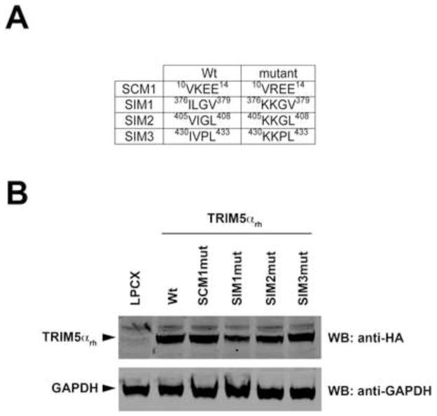
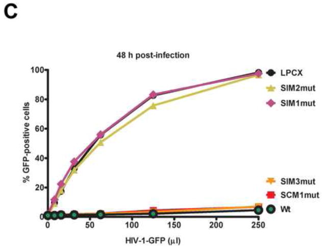
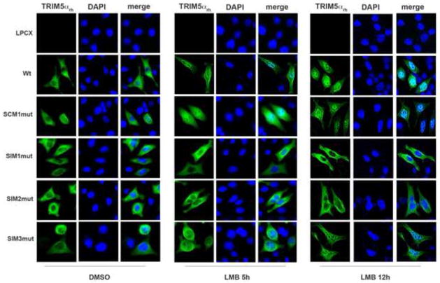
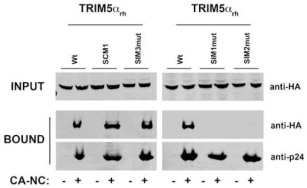
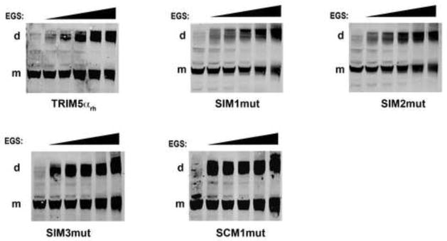
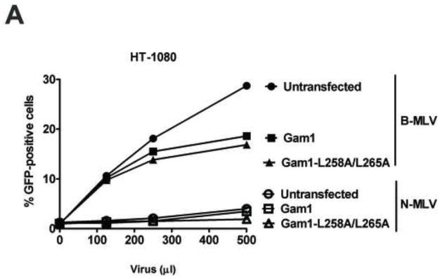
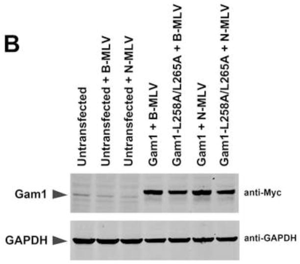
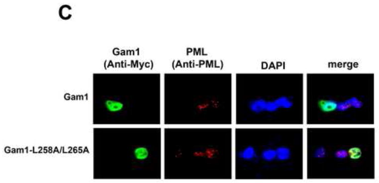
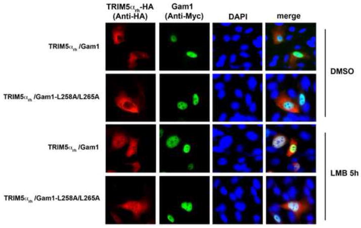
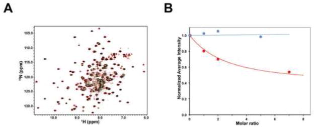
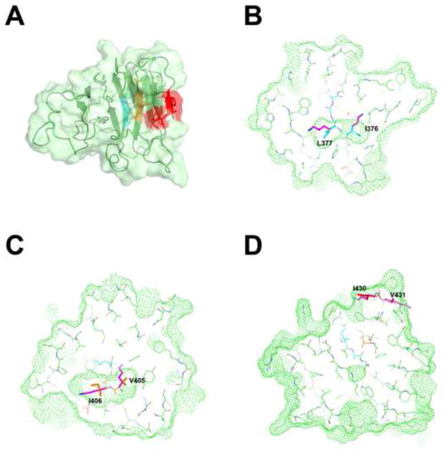
Recent findings suggested that the SUMO-interacting motifs (SIMs) present in the human TRIM5α (TRIM5α(hu)) protein play an important role in the ability of TRIM5α(hu) to restrict N-MLV. Here we explored the role of SIMs in the ability of rhesus TRIM5α (TRIM5α(rh)) to restrict HIV-1, and found that TRIM5α(rh) SIM mutants IL376KK (SIM1mut) and VI405KK (SIM2mut) completely lost their ability to block HIV-1 infection. Interestingly, these mutants also lost the recently described property of TRIM5α(rh) to shuttle into the nucleus. Analysis of these variants revealed that they are unable to interact with the HIV-1 core, which might explain the reason that these variants are not active against HIV-1. Furthermore, NMR titration experiments to assay the binding between the PRYSPRY domain of TRIM5α(rh) and the small ubiquitin-like modifier 1(SUMO-1) revealed no interaction. In addition, we examined the role of SUMOylation in restriction, and find out that inhibition of SUMOylation by the adenoviral protein Gam1 did not alter the retroviral restriction ability of TRIM5α. Overall, our results do not support a role for SIMs or SUMOylation in the antiviral properties of TRIM5α.


Figures

Figure 1
Figure 1. Contribution of TRIM5αrh SUMO-conjugation and SUMO-interacting motifs(SIM) to restriction of HIV-1
Dog Cf2Th cells were transduced with the LPCX vector expressing HA-tagged wild-type and mutant TRIM5αrh proteins. The TRIM5αrh SUMO-conjugation motif(SCM1) and the three SUMO-interacting motifs (SIM1, SIM2 and SIM3) variants are shown with their respective amino acid position (A). Stable cell lines were selected with 5 μg/ml puromycin, and the expression levels of mutant and wild-type TRIM5αrh proteins were assayed by Western blotting using anti-HA and anti-GAPDH antibodies (B). Stable cell lines were challenged with different amounts of HIV-1-GFP (C). The percentage of GFP-positive cells was measured 48 h later by FACS. The results of three independent experiments were similar; the results of a single experiment are shown.
Figure 2
Figure 2. Intracellular distribution of TRIM5αrh SIM mutant proteins in the presence of leptomycin B
Human HeLa cells expressing the indicated TRIM5αrh variants were treated with 20 ng/ml of leptomycin B (LMB) or DMSO for either 5 or 12 h. Treated cells were were fixed and immunostained using anti-HA antibodies (green). Cellular nuclei were stained by using DAPI (blue). Representative figures are shown. The results of three independent experiments were similar (Supplemental Table 1); the result of a single experiment is shown.
Figure 3
Figure 3. Binding of TRIM5αrh SIM mutant proteins to in vitro assembled HIV-1 capsid-nucleocapsid (CA-NC) complexes
Human 293T cells were transfected with plasmids expressing the indicated wild-type and mutant HA-tagged TRIM5αrh proteins. Forty-eight h after transfection, cells were lysed. The lysates were incubated at room temperature for 1 h with in vitro assembled HIV-1 CA-NC complexes. The mixtures were applied to a 70% sucrose cushion and centrifuged. INPUT represents the lysates analyzed by Western blotting before being applied to the 70% cushion. The input mixtures were Western blotted using anti-HA antibodies. Similarly, the pellets from the 70% cushion (BOUND) were analyzed by Western blotting using anti-HA and anti-p24 antibodies. The results of three independent experiments were similar; the result of a single experiment is shown.
Figure 4
Figure 4. Oligomerization of TRIM5αrh SIM mutant proteins
Lysates from 293T cells expressing wild-type and mutant TRIM5αrh proteins were cross-linked with increasing concentrations of ethylene glycol-bis(succinimidyl succinate) (EGS), and were subjected to Western blotting using anti-HA antibodies, as described in Materials and Methods. “m” and “d” stands for monomer and dimer, respectively. Similar results were obtained in three independent experiments and a representative experiment is shown.
Figure 5
Figure 5. Role of SUMOylation in the ability of TRIM5αhu to restrict N-MLV
Human HT-1080 cells were transienly transfected with plasmids expressing Myc-tagged Gam1 or Gam1-L258A/L265A. After 24 h, cells were challenged with increasing amounts of N-MLV-GFP or B-MLV-GFP. Viruses expressing the reproter GFP were normilized in dog Cf2Th cells. Forthy-eight h post-transfection, infection was determined by counting the percentage of GFP-positive cells using FACS (A). The expression of Gam1 and Gam1-L258A/L265A were analized by Western blotting using anti-Myc and anti-GAPDH antibodies (B). The ability of Gam1 to destroy PML-containing nuclear bodies was assayed by transiently transfecting HT-1080 cells with plasmids expressing Gam1 or Gam1-L258A/L265A tagged with a Myc epitope. After 24 h, cells were fixed and and immunostained using anti-Myc (green) and anti-PML (red) antibodies (C). Cellular nuclei were stained by using DAPI (blue). Similar results were obtained in three independent experiments and a representative experiment is shown.
Figure 6
Figure 6. Inhibition of SUMOylation by Gam1 does not affect the ability of TRIM5αrh to traffic into the nucleus
Human HT-1080 cells were cotransfected with plasmids expressing Myc-tagged Gam1 and HA-tagged TRIM5αrh. After 24 h, cells were treated with 20 ng/ml of leptomycin B (LMB) or DMSO for 5 h, fixed and immunostained using antibodies anit-myc (green) and anti-HA (red) antibodies. Cellular nuclei were stained by using DAPI (blue). Similar results were obtained in three independent experients(Supplemental Table 2), and a representative experiment is shown.
Figure 7
Figure 7. NMR titrations of the TRIM5αrh PRYSPRY domain with SUMO-1
(A) The superposition of the 15N-HSQC spectra of the 0.3 mM 15N-labeled PRYSPRY sample in the presence of 1.5 mM SUMO-1 (green) or in the absence of SUMO-1 added (red) is shown. Overlapping signals appear black. (B) NMR titrations of the 15N-labeled PRYSPRY domain with SUMO-1 (blue) and the N-terminal domain of HIV-1 capsid (red). Addition of 5:1 molar excess of SUMO-1 does not show any detectable attenuation of the PRYSPRY NMR signals.
Figure 8
Figure 8. Putative SUMO interaction motifs (SIM) mapped onto the TRIM5αrh PRYSPRY structure
(A)SIM1, SIM2 and SIM3 mapped onto the TRIM5αrh PRYSPRY structure. The SPRY domain is shown as a cartoon with the protein surface colored in light green. The segments of the protein identified as SUMO interacting motifs SIM-1, SIM-2 and SIM-3 are colored in cyan, orange and red respectively. Only the SIM-3 segment contains surface-exposed residues, while SIM-1 and SIM-2 are buried in the hydrophobic core of the protein. (B) Slice through the TRIM5αrh PRYSPRY structure showing the volume (green mesh) occupied by residues I376 and L377 of SIM1 (cyan). The two residues are completely buried within the hydrophobic core and the lysine residues of the SIM1 mutant (magenta) can not be accomodated within that space. The resulting steric clash and buried positive charge are incompatible with the stably-folded native structure. (C) The same is true for SIM2. (D) In contrast, SIM3 is exposed on the protein surface, therefore SIM3mut is not expected to disrupt the overall PRYSPRY structure.

Similar articles

Cited by

References

    1. Arriagada G, Muntean LN, Goff SP. SUMO-interacting motifs of human TRIM5alpha are important for antiviral activity. PLoS Pathog. 2011;7:e1002019. - PMC - PubMed
    1. Biris N, Yang Y, Taylor AB, Tomashevski A, Guo M, Hart PJ, Diaz-Griffero F, Ivanov DN. Structure of the rhesus monkey TRIM5alpha PRYSPRY domain, the HIV capsid recognition module. Proc Natl Acad Sci U S A 2012 - PMC - PubMed
    1. Boggio R, Colombo R, Hay RT, Draetta GF, Chiocca S. A mechanism for inhibiting the SUMO pathway. Mol Cell. 2004;16:549–561. - PubMed
    1. Boggio R, Passafaro A, Chiocca S. Targeting SUMO E1 to ubiquitin ligases: a viral strategy to counteract sumoylation. J Biol Chem. 2007;282:15376–15382. - PubMed
    1. Chiocca S, Baker A, Cotten M. Identification of a novel antiapoptotic protein, GAM-1, encoded by the CELO adenovirus. J Virol. 1997;71:3168–3177. - PMC - PubMed

Publication types

MeSH terms

LinkOut - more resources