

Kinematic differences between optical motion capture and biplanar videoradiography during a jump-cut maneuver
- PMID: 23084785
- PMCID: PMC3551998
- DOI: 10.1016/j.jbiomech.2012.09.023
Kinematic differences between optical motion capture and biplanar videoradiography during a jump-cut maneuver
Abstract
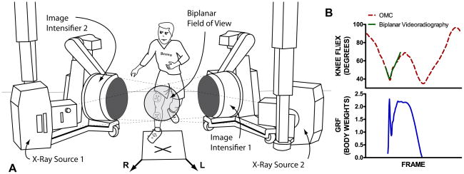
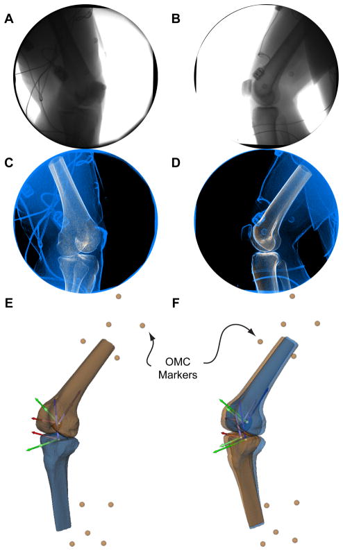
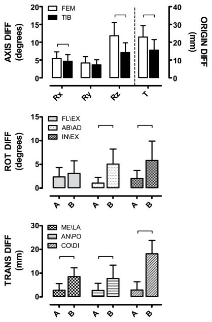
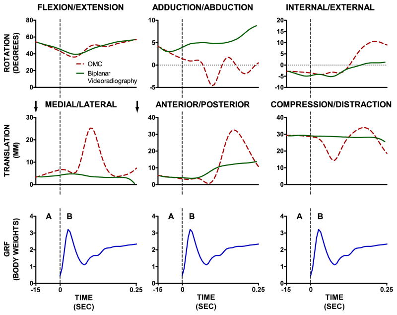
Jumping and cutting activities are investigated in many laboratories attempting to better understand the biomechanics associated with non-contact ACL injury. Optical motion capture is widely used; however, it is subject to soft tissue artifact (STA). Biplanar videoradiography offers a unique approach to collecting skeletal motion without STA. The goal of this study was to compare how STA affects the six-degrees-of-freedom motion of the femur and tibia during a jump-cut maneuver associated with non-contact ACL injury. Ten volunteers performed a jump-cut maneuver while their landing leg was imaged using optical motion capture (OMC) and biplanar videoradiography. The within-bone motion differences were compared using anatomical coordinate systems for the femur and tibia, respectively. The knee joint kinematic measurements were compared during two periods: before and after ground contact. Over the entire activity, the within-bone motion differences between the two motion capture techniques were significantly lower for the tibia than the femur for two of the rotational axes (flexion/extension, internal/external) and the origin. The OMC and biplanar videoradiography knee joint kinematics were in best agreement before landing. Kinematic deviations between the two techniques increased significantly after contact. This study provides information on the kinematic discrepancies between OMC and biplanar videoradiography that can be used to optimize methods employing both technologies for studying dynamic in vivo knee kinematics and kinetics during a jump-cut maneuver.
Copyright © 2012 Elsevier Ltd. All rights reserved.
Conflict of interest statement
The authors have no financial or personal relationships that could bias this work.
Figures
References
-
- Akbarshahi M, Schache AG, Fernandez JW, Baker R, Banks S, Pandy MG. Non-invasive assessment of soft-tissue artifact and its effect on knee joint kinematics during functional activity. Journal of Biomechics. 2010;43:1292–1301. - PubMed
-
- Andriacchi TP, Alexander EJ, Toney MK, Dyrby C, Sum J. A point cluster method for in vivo motion analysis: applied to a study of knee kinematics. Journal of Biomechanical Engineering. 1998;120:743–749. - PubMed
-
- Boden BP, Dean GS, Feagin JA, Garrett WE. Mechanisms of anterior cruciate ligament injury. Orthopedics. 2000;23:573–578. - PubMed
-
- Brainerd EL, Baier DB, Gatesy SM, Hedrick TL, Metzger KA, Gilbert SL, Crisco JJ. X-ray reconstruction of moving morphology (XROMM): precision, accuracy and applications in comparative biomechanics research. Journal of Experimental Zoology Part A, Ecological Genetics and Physiology. 2010;313:262–279. - PubMed
-
- Buczek FL, Rainbow MJ, Cooney KM, Walker MR, Sanders JO. Implications of using hierarchical and six degree-of-freedom models for normal gait analyses. Gait & Posture. 2010;31:57–63. - PubMed
Publication types
MeSH terms
Grants and funding
LinkOut - more resources
Full Text Sources
Other Literature Sources
Medical
