

Human conjunctival microvasculature assessed with a retinal function imager (RFI)
- PMID: 23084966
- PMCID: PMC3534915
- DOI: 10.1016/j.mvr.2012.10.003
Human conjunctival microvasculature assessed with a retinal function imager (RFI)
Abstract
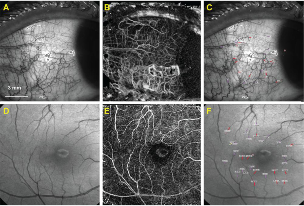
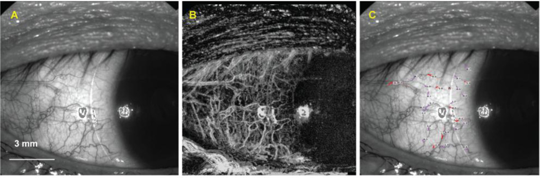
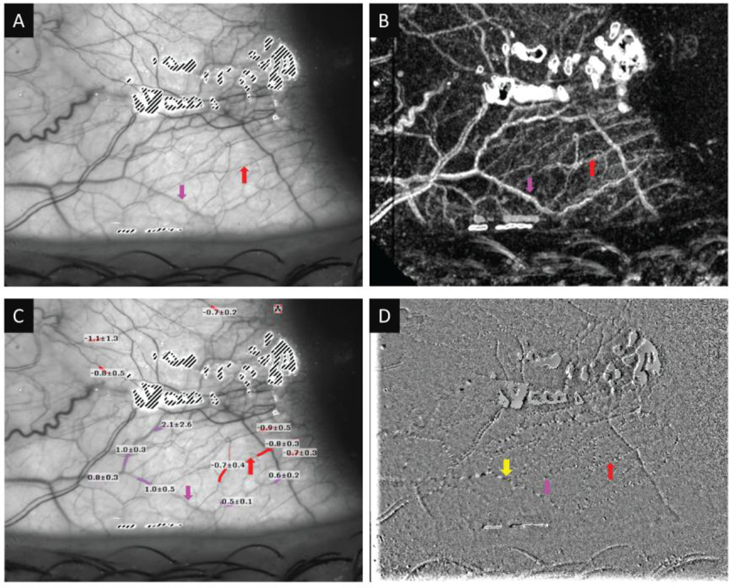
The conjunctival and cerebral vasculatures share similar embryological origins, with similar structural and physiological characteristics. Tracking the conjunctival microvasculature may provide useful information for predicting the onset, progression and prognosis of both systemic and central nervous system (CNS) vascular diseases. The bulbar conjunctival vasculature was imaged using a retinal function imager (RFI, Optical Imaging Ltd, Rehovot, Israel). Hemoglobin in red blood cells was used as an intrinsic motion-contrast agent in the generation of detailed noninvasive capillary-perfusion maps (nCPMs) and the calculation of the blood flow velocity. Five healthy subjects were imaged under normal conditions and again under the stress condition of wearing a contact lens. The retina was also imaged in one eye of one subject for comparison. The nCPMs showed the conjunctival microvasculature in exquisite detail, which appeared as clear as the retinal nCPMs. The blood flow velocities in the temporal conjunctival microvasculature were 0.86±0.08 (mean±SD, mm/s) for the bare eye and 0.99±0.11 mm/s with contact lens wear. It is feasible to use RFI for imaging the conjunctival vasculature.
Copyright © 2012 Elsevier Inc. All rights reserved.
Figures
Similar articles
-
Altered Bulbar Conjunctival Microcirculation in Response to Contact Lens Wear.Eye Contact Lens. 2017 Mar;43(2):95-99. doi: 10.1097/ICL.0000000000000241. Eye Contact Lens. 2017. PMID: 27078615 Free PMC article.
-
Functional slit lamp biomicroscopy for imaging bulbar conjunctival microvasculature in contact lens wearers.Microvasc Res. 2014 Mar;92:62-71. doi: 10.1016/j.mvr.2014.01.005. Epub 2014 Jan 17. Microvasc Res. 2014. PMID: 24444784 Free PMC article.
-
Changes in bulbar conjunctival microcirculation and microvasculature during short-term scleral lens wearing and their associated factors.Cont Lens Anterior Eye. 2024 Oct;47(5):102159. doi: 10.1016/j.clae.2024.102159. Epub 2024 Jun 8. Cont Lens Anterior Eye. 2024. PMID: 38851944
-
Functional imaging using the retinal function imager: direct imaging of blood velocity, achieving fluorescein angiography-like images without any contrast agent, qualitative oximetry, and functional metabolic signals.Jpn J Ophthalmol. 2009 Jul;53(4):345-51. doi: 10.1007/s10384-009-0689-0. Epub 2009 Sep 8. Jpn J Ophthalmol. 2009. PMID: 19763751 Review.
-
A Mini Review of Clinical and Research Applications of the Retinal Function Imager.Curr Eye Res. 2018 Mar;43(3):273-288. doi: 10.1080/02713683.2017.1414853. Epub 2018 Jan 8. Curr Eye Res. 2018. PMID: 29308926 Review.
Cited by
-
Paradigm Shifts in Ophthalmic Diagnostics.Trans Am Ophthalmol Soc. 2016 Aug;114:WP1. Trans Am Ophthalmol Soc. 2016. PMID: 28008209 Free PMC article. Review.
-
Altered Bulbar Conjunctival Microcirculation in Response to Contact Lens Wear.Eye Contact Lens. 2017 Mar;43(2):95-99. doi: 10.1097/ICL.0000000000000241. Eye Contact Lens. 2017. PMID: 27078615 Free PMC article.
-
A novel algorithm for cardiovascular screening using conjunctival microcirculatory parameters and blood biomarkers.Sci Rep. 2022 Apr 21;12(1):6545. doi: 10.1038/s41598-022-10491-7. Sci Rep. 2022. PMID: 35449196 Free PMC article.
-
Automated Assessment of Hemodynamics in the Conjunctival Microvasculature Network.IEEE Trans Med Imaging. 2016 Feb;35(2):605-11. doi: 10.1109/TMI.2015.2486619. Epub 2015 Oct 6. IEEE Trans Med Imaging. 2016. PMID: 26452274 Free PMC article.
-
Automated Real-Time Conjunctival Microvasculature Image Stabilization.IEEE Trans Med Imaging. 2016 Jul;35(7):1670-5. doi: 10.1109/TMI.2016.2522918. Epub 2016 Feb 3. IEEE Trans Med Imaging. 2016. PMID: 26863649 Free PMC article.
References
-
- Beutelspacher SC, Serbecic N, Barash H, Burgansky-Eliash Z, Grinvald A, Jonas JB. Central serous chorioretinopathy shows reduced retinal flow circulation in retinal function imaging (RFI) Acta Ophthalmol. 2011;89:e479–e482. - PubMed
-
- Cheung AT, Chan MS, Ramanujam S, Rangaswami A, Curl K, Franklin P, Wun T. Effects of poloxamer 188 treatment on sickle cell vaso-occlusive crisis: computer-assisted intravital microscopy study. J. Investig. Med. 2004;52:402–406. - PubMed
-
- Cheung AT, Chen PC, Larkin EC, Duong PL, Ramanujam S, Tablin F, Wun T. Microvascular abnormalities in sickle cell disease: a computer-assisted intravital microscopy study. Blood. 2002a;99:3999–4005. - PubMed
-
- Cheung AT, Chen PC, Leshchinsky TV, Wiltse SL, Basadonna GP, Katznelson S, Perez RV. Improvement in conjunctival microangiopathy after simultaneous pancreas-kidney transplants. Transplant. Proc. 1997;29:660–661. - PubMed
-
- Cheung AT, Harmatz P, Wun T, Chen PC, Larkin EC, Adams RJ, Vichinsky EP. Correlation of abnormal intracranial vessel velocity, measured by transcranial Doppler ultrasonography, with abnormal conjunctival vessel velocity, measured by computer-assisted intravital microscopy, in sickle cell disease. Blood. 2001a;97:3401–3404. - PubMed
MeSH terms
Substances
Grants and funding
LinkOut - more resources
Full Text Sources
Other Literature Sources
