

Rapid whisker movements in sleeping newborn rats
- PMID: 23084988
- PMCID: PMC3494768
- DOI: 10.1016/j.cub.2012.09.009
Rapid whisker movements in sleeping newborn rats
Abstract
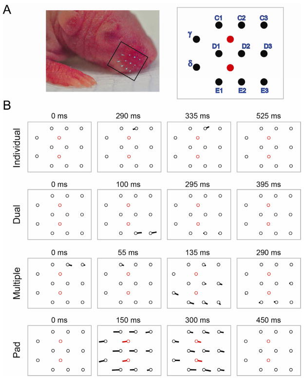
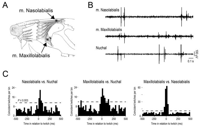
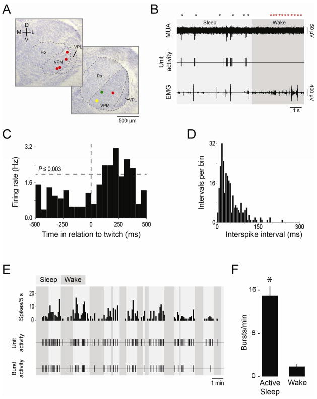
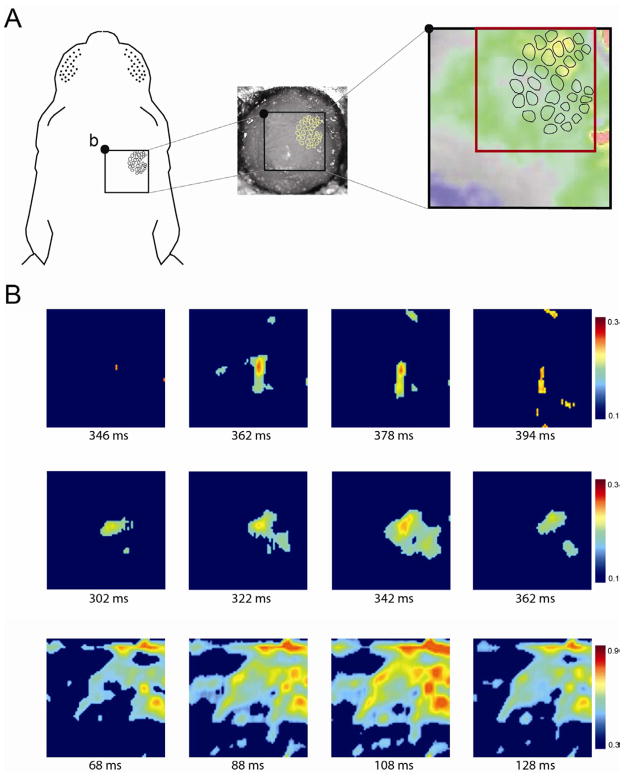
Spontaneous activity in the sensory periphery drives infant brain activity and is thought to contribute to the formation of retinotopic and somatotopic maps. In infant rats during active (or REM) sleep, brainstem-generated spontaneous activity triggers hundreds of thousands of skeletal muscle twitches each day; sensory feedback from the resulting limb movements is a primary activator of forebrain activity. The rodent whisker system, with its precise isomorphic mapping of individual whiskers to discrete brain areas, has been a key contributor to our understanding of somatotopic maps and developmental plasticity. But although whisker movements are controlled by dedicated skeletal muscles, spontaneous whisker activity has not been entertained as a contributing factor to the development of this system. Here we report in 3- to 6-day-old rats that whiskers twitch rapidly and asynchronously during active sleep; furthermore, neurons in whisker thalamus exhibit bursts of activity that are tightly associated with twitches but occur infrequently during waking. Finally, we observed barrel-specific cortical activity during periods of twitching. This is the first report of self-generated, sleep-related twitches in the developing whisker system, a sensorimotor system that is unique for the precision with which it can be experimentally manipulated. The discovery of whisker twitching will allow us to attain a better understanding of the contributions of peripheral sensory activity to somatosensory integration and plasticity in the developing nervous system.
Copyright © 2012 Elsevier Ltd. All rights reserved.
Figures
Similar articles
-
The Nature of the Sensory Input to the Neonatal Rat Barrel Cortex.J Neurosci. 2016 Sep 21;36(38):9922-32. doi: 10.1523/JNEUROSCI.1781-16.2016. J Neurosci. 2016. PMID: 27656029 Free PMC article.
-
Self-Generated Whisker Movements Drive State-Dependent Sensory Input to Developing Barrel Cortex.Curr Biol. 2020 Jun 22;30(12):2404-2410.e4. doi: 10.1016/j.cub.2020.04.045. Epub 2020 May 14. Curr Biol. 2020. PMID: 32413304 Free PMC article.
-
Modelling the emergence of whisker barrels.Elife. 2020 Sep 29;9:e55588. doi: 10.7554/eLife.55588. Elife. 2020. PMID: 32988453 Free PMC article.
-
Cortical control of whisker movement.Annu Rev Neurosci. 2014;37:183-203. doi: 10.1146/annurev-neuro-062012-170344. Epub 2014 May 9. Annu Rev Neurosci. 2014. PMID: 24821429 Review.
-
Neuronal Circuits in Barrel Cortex for Whisker Sensory Perception.Physiol Rev. 2021 Jan 1;101(1):353-415. doi: 10.1152/physrev.00019.2019. Epub 2020 Aug 20. Physiol Rev. 2021. PMID: 32816652 Review.
Cited by
-
Early brain activity: Translations between bedside and laboratory.Prog Neurobiol. 2022 Jun;213:102268. doi: 10.1016/j.pneurobio.2022.102268. Epub 2022 Mar 29. Prog Neurobiol. 2022. PMID: 35364141 Free PMC article. Review.
-
Discharge and Role of Acetylcholine Pontomesencephalic Neurons in Cortical Activity and Sleep-Wake States Examined by Optogenetics and Juxtacellular Recording in Mice.eNeuro. 2018 Sep 13;5(4):ENEURO.0270-18.2018. doi: 10.1523/ENEURO.0270-18.2018. eCollection 2018 Jul-Aug. eNeuro. 2018. PMID: 30225352 Free PMC article.
-
Developmental features of sleep electrophysiology in family dogs.Sci Rep. 2021 Nov 23;11(1):22760. doi: 10.1038/s41598-021-02117-1. Sci Rep. 2021. PMID: 34815446 Free PMC article.
-
A new view of "dream enactment" in REM sleep behavior disorder.Sleep Med Rev. 2016 Dec;30:34-42. doi: 10.1016/j.smrv.2015.12.002. Epub 2015 Dec 17. Sleep Med Rev. 2016. PMID: 26802823 Free PMC article. Review.
-
The Nature of the Sensory Input to the Neonatal Rat Barrel Cortex.J Neurosci. 2016 Sep 21;36(38):9922-32. doi: 10.1523/JNEUROSCI.1781-16.2016. J Neurosci. 2016. PMID: 27656029 Free PMC article.
References
Publication types
MeSH terms
Grants and funding
LinkOut - more resources
Full Text Sources
