. 2012 Nov 20;22(22):2104-13.
doi: 10.1016/j.cub.2012.09.018. Epub 2012 Oct 18.

Lipid droplets control the maternal histone supply of Drosophila embryos

Affiliations

Lipid droplets control the maternal histone supply of Drosophila embryos

Zhihuan Li et al. Curr Biol. .

Abstract

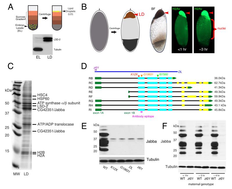
Background: Histones are essential for chromatin packing, yet free histones not incorporated into chromatin are toxic. While in most cells multiple regulatory mechanisms prevent accumulation of excess histones, early Drosophila embryos contain massive extranuclear histone stores, thought to be essential for development. Excess histones H2A, H2B, and H2Av are bound to lipid droplets, ubiquitous fat storage organelles especially abundant in embryos. It has been proposed that sequestration on lipid droplets allows safe transient storage of supernumerary histones.

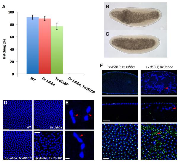
Results: Here, we critically test this sequestration hypothesis. We find that histones are anchored to lipid droplets via the previously uncharacterized protein Jabba: Jabba localizes to droplets, coimmunoprecipitates with histones, and is necessary to recruit histones to droplets. Jabba mutants lack the maternal H2A, H2B, and H2Av deposits altogether; presumably, these deposits are eliminated unless sequestered on droplets. Jabba mutant embryos compensate for this histone deficit by translating maternal histone mRNAs. However, when histone expression is mildly compromised, the maternal histone protein deposits are essential for proper early mitoses and for viability.

Conclusions: A growing number of proteins from other cellular compartments have been found to transiently associate with lipid droplets. Our studies provide the first insight into mechanism and functional relevance of this sequestration. We conclude that sequestration on lipid droplets allows embryos to build up extranuclear histone stores and provides histones for chromatin assembly during times of high demand. This work reveals a novel aspect of histone metabolism and establishes lipid droplets as functional storage sites for unstable or detrimental proteins.


Figures

Figure 1
Figure 1
Identification and characterization of Jabba. (A) Purification of lipid droplets from embryo lysate (EL) by floatation. During centrifugation, lipid droplets float to the top of the sucrose gradient. The lipid-droplet fraction (LD) is enriched for the droplet protein LSD-2 and depleted for the cytoplasmic protein tubulin. (B) Enrichment of lipid droplets by in-vivo centrifugation. The droplets form a distinct lipid-droplet layer (LD; arrowheads), visible by bright-field (BF) microscopy. Anti-H2Av staining reveals H2Av on the droplet layer of embryos of different ages, as well as within nuclei. (C) Proteins from purified wild-type droplets were separated by SDS PAGE and stained with Coomassie Blue (MW = molecular weight markers, LD = lipid-droplet fraction). Major bands were excised and identified by mass spectrometry. (D) Jabba locus. Exons are shown as boxes: green = UTR, cyan = coding regions shared between all isoforms, yellow = coding regions restricted to some isoforms. Pink bar = peptide used for antibody generation. Extent of deletions in JabbaDL and Jabbazl01 is indicated as well as the location of the non-sense mutation JabbaK12X and of two transposable element insertions. (E) Lysates from ~3 hr old embryos analyzed by anti-Jabba immunoblotting. (F) Embryos from reciprocal crosses between wild-type and Jabbazl01 parents analyzed by anti-Jabba immunoblotting. See also Fig. S1.
Figure 2
Figure 2
Jabba is a lipid-droplet protein. (A) Equal amounts of protein from wild-type lipid-droplet samples (LD) and embryo lysate (EL) analyzed by immunoblotting (see Fig. 1A). Two Jabba bands are highly enriched in the droplet fraction; under these conditions, no signal is detected in the lysate. (B) Embryos expressing the droplet marker GFP-LD stained for GFP (green) and Jabba (red). Jabba is present in rings that colocalize with GFP-LD. Scale bar = 2.5 μm. (C) Anti-Jabba staining of centrifuged wild-type and Jabbazl01 embryos. Jabba is highly enriched in the droplet layer (arrowhead) in the wild type. The middle panel shows a ~10 fold longer exposure of the mutant. (D) Anti-Jabba staining of centrifuged egg chambers. Lipid-droplet layers in the oocyte (arrow) and in nurse cells (arrowhead) are indicated. (E) Kc167 cells transiently expressing Jabba-PB-eGFP (green) stained with LipidTOX (red) to reveal lipid droplets. Scale bar = 5 μm. See also Fig. S2.
Figure 3
Figure 3
Jabba mutants have abundant lipid droplets with altered protein content. (A) Triglyceride levels in 2 hr old wild-type and Jabbazl01 embryos are similar. Error bars represent standard deviations. (B) Lipid droplets in 2 hr old wild-type and Jabbazl01 embryos, revealed by Nile Red staining. Jabbazl01 droplets are unevenly distributed and irregularly shaped, a phenotype that inspired the name Jabba. Scale bar = 5 μm. (C) Equal amounts of proteins of droplets purified from wild-type and Jabbazl01 embryos were separated by SDS PAGE and stained with Coomassie Blue.
Figure 4
Figure 4
Jabba is necessary for histone recruitment to lipid droplets. (A) Immunoblotting of purified droplets from wild-type and Jabbazl01 embryos. Equal amounts of total protein were loaded. Kinesin heavy chain (KHC) levels are similar. H2A, H2B, and H2Av levels are dramatically reduced in the mutant. (B) Immunostaining of centrifuged embryos (<1 hr). Jabbazl01 embryos lack H2A, H2B, and H2Av signal in the droplet layer (top). (C) Centrifuged embryos from mothers expressing H2Av-GFP, by bright-field and fluorescence microscopy. GFP signal on the droplet layer (arrowhead) is dramatically reduced in the mutant. (D) In uncentrifuged embryos, H2Av-GFP is present in nuclei for both genotypes, but is absent from cytoplasmic puncta (which represent lipid droplets [15]) in Jabbazl01. The exposure for the wild type is double that of the mutant sample; for unknown reasons, H2Av-GFP signal in the nuclei is stronger in Jabbazl01 embryos. Scale bars: 7.5 μm (top), 5 μm (bottom). See also Fig. S4.
Figure 5
Figure 5
Jabba is the histone docking protein on droplets. (A) Anti-Jabba immunoblotting in equal numbers of embryos from mothers carrying two, one, or zero copies of a wild-type Jabba gene. Jabba protein levels scale with Jabba gene dosage (2x Jabba = wild type; 1x Jabba = Df(2R)Exel7158/+; 0x Jabba = Jabbazl01). (B) H2B signal on the lipid-droplet layers of centrifuged 2x Jabba embryos (~3 hrs old) is much stronger than that of 1x Jabba, while nuclear signal is similar. (C) Centrifuged H2Av-GFP/+ and Jabbazl01/+, H2Av-GFP/+ embryos. Embryo ages: <1 hr (left panel), ~3 hrs (right panel). Reduced droplet signal with age presumably represents transfer of histones from lipid droplets to nuclei [15]. (D) H2Av-GFP co-immunoprecipitates with Jabba. Lipid droplets purified from H2Av-GFP and wild-type embryos were exposed to anti-GFP antibodies for immunoprecipitation.
Figure 6
Figure 6
Jabba mutant embryos lack maternal histone deposits, but can synthesize histones zygotically. (A,B) Compared to wild type, unfertilized Jabbazl01 embryos have barely detectable H2A and H2B, less H2Av, but similar amounts of H3. (C) By immunoblotting, the amount of H2A and H2B in unfertilized 2x Jabba embryos is roughly double that of 1x Jabba embryos. (D) Jabbazl01 embryos can generate H2A and H2B. Levels of both histones go up dramatically as development proceeds (stage 1 ~ 30 min; stage 2 ~ 60 min; stage 4 ~ 2 hrs; stage 5 ~ 3 hrs). (E) By ~3 hrs, Jabbazl01 embryos have reached similar H2B levels as wild type. (F) Unfertilized wild-type and Jabbazl01 embryos have similar levels of H2A mRNA (measured by qRT-PCR, normalized to the wild-type value). Error bars represent standard deviations.
Figure 7
Figure 7
Reduced histone expression and Jabba mutants are synthetically lethal. (A) Hatching frequency of embryos from mothers of various genotypes. 0x Jabba = Jabbazl01; 1x dSLBP = heterozygous for Df(3R)3450. Error bars represent standard deviations. (B, C) Late syncytial blastoderm embryos (heat-fixed and inspected by DIC microscopy) from 1x dSLBP 0x Jabba mothers, with aberrant yolk distribution (B) or grossly normal morphology (C). By cellularization, most embryos displayed some morphological defects. (D) Surface view of blastoderm embryos; genotype of mothers is indicated (blue = DNA; scale bar = 25 μm) (E) Nuclei of an embryo from a 1x dSLBP 0x Jabba mother showing chromosomes (arrowheads) connecting daughter nuclei late in mitosis. Scale bar = 5 μm. (F) Blastoderm embryos stained for DNA (blue) or centrosomes (green): cross sections at low (top) and high (middle) magnification, plus a surface view (bottom). In the absence of Jabba, nuclei are absent from cortical patches (arrowhead, bottom) and present between the cortex and the central yolk (arrowheads, top and middle). Scale bars = 10 μm. See also Fig. S5 and Movie S6.

Comment in

References

    1. An W, Leuba SH, van Holde K, Zlatanova J. Linker histone protects linker DNA on only one side of the core particle and in a sequence-dependent manner. Proc Natl Acad Sci U S A. 1998;95:3396–3401. - PMC - PubMed
    1. Celona B, Weiner A, Di Felice F, Mancuso FM, Cesarini E, Rossi RL, Gregory L, Baban D, Rossetti G, Grianti P, et al. Substantial histone reduction modulates genomewide nucleosomal occupancy and global transcriptional output. PLoS Biol. 2011;9:e1001086. - PMC - PubMed
    1. O’Sullivan RJ, Kubicek S, Schreiber SL, Karlseder J. Reduced histone biosynthesis and chromatin changes arising from a damage signal at telomeres. Nat Struct Mol Biol. 2010;17:1218–1225. - PMC - PubMed
    1. Feser J, Truong D, Das C, Carson JJ, Kieft J, Harkness T, Tyler JK. Elevated histone expression promotes life span extension. Mol Cell. 2010;39:724–735. - PMC - PubMed
    1. Gunjan A, Verreault A. A Rad53 kinase-dependent surveillance mechanism that regulates histone protein levels in S. cerevisiae. Cell. 2003;115:537–549. - PubMed

Publication types