

Distinct requirements for Sin3a in perinatal male gonocytes and differentiating spermatogonia
- PMID: 23085237
- PMCID: PMC3508146
- DOI: 10.1016/j.ydbio.2012.10.009
Distinct requirements for Sin3a in perinatal male gonocytes and differentiating spermatogonia
Abstract
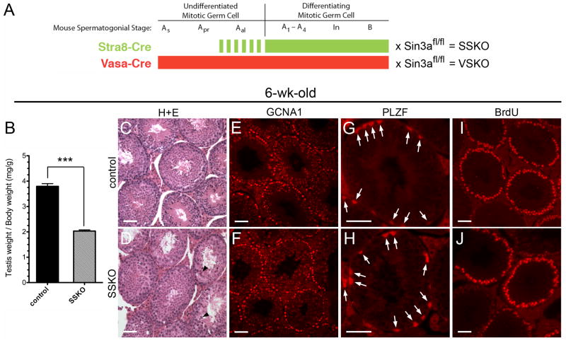
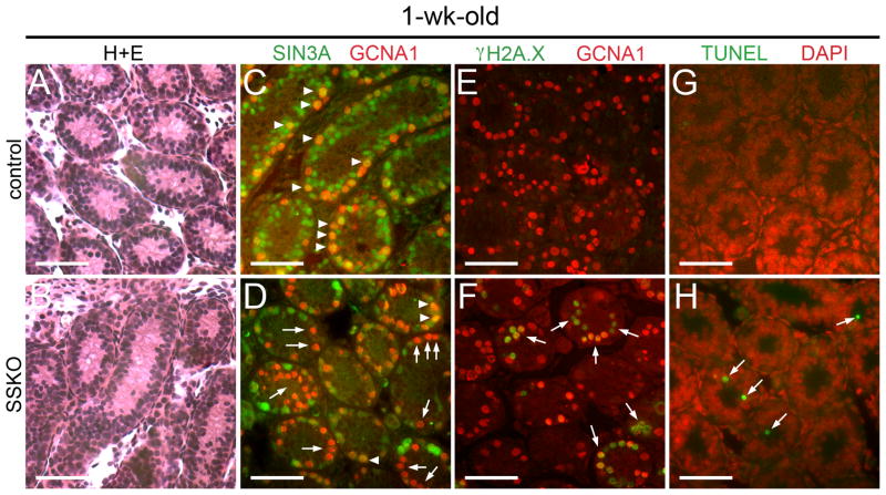
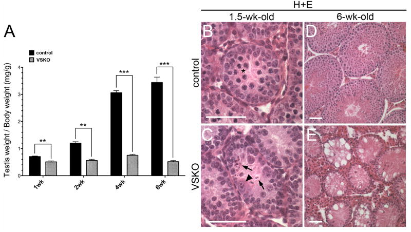
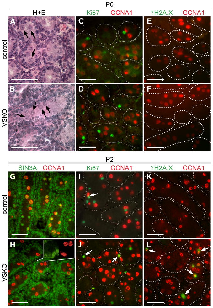
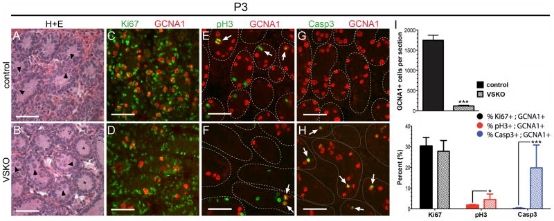
Chromatin modifier Swi-independent 3a (SIN3A), together with associated histone deacetylases, influences gene expression during development and differentiation through a variety of transcription factors in a cell-specific manner. Sin3a is essential for the maintenance of inner cell mass cells of mouse blastocysts, embryonic fibroblasts, and myoblasts, but is not required for the survival of trophectoderm or Sertoli cells. To better understand how this transcriptional regulator modulates cells at different developmental stages within a single lineage, we used conditional gene targeting in mice to ablate Sin3a from perinatal quiescent male gonocytes and from postnatal differentiating spermatogonia. Mitotic germ cells expressing stimulated by retinoic acid gene 8 (Stra8) that lacked Sin3a exhibited increased DNA damage and apoptosis, yet collectively progressed through meiosis and spermiogenesis and generated epididymal sperm at approximately 50% of control levels, sufficient for normal fertility. In contrast, perinatal gonocytes lacking Sin3a underwent rapid depletion that coincided with cell cycle reentry, exhibiting 2.5-fold increased histone H3 phosphorylation upon cycling that suggested a prophase/metaphase block; germ cells were almost entirely absent two weeks after birth, resulting in sterility. Gene expression profiling of neonatal testes containing Sin3a-deleted gonocytes identified upregulated transcripts highly associated with developmental processes and pattern formation, and downregulated transcripts involved in nuclear receptor activity, including Nr4a1 (Nur77). Interestingly, Nr4a1 levels were elevated in testes containing Stra8-expressing, Sin3a-deleted spermatogonia. SIN3A directly binds to the Nr4a1 promoter, and Nr4a1 expression is diminished upon spermatogonial differentiation in vitro. We conclude that within the male germline, Sin3a is required for the mitotic reentry of gonocytes, but is dispensable for the maintenance of differentiating spermatogonia and subsequent spermatogenic processes.
Copyright © 2012 Elsevier Inc. All rights reserved.
Conflict of interest statement
Figures

Similar articles
-
Chromatin associated Sin3A is essential for male germ cell lineage in the mouse.Dev Biol. 2012 Sep 15;369(2):349-55. doi: 10.1016/j.ydbio.2012.07.006. Epub 2012 Jul 20. Dev Biol. 2012. PMID: 22820070 Free PMC article.
-
Sin3a is required by sertoli cells to establish a niche for undifferentiated spermatogonia, germ cell tumors, and spermatid elongation.Stem Cells. 2010 Aug;28(8):1424-34. doi: 10.1002/stem.464. Stem Cells. 2010. PMID: 20572009 Free PMC article.
-
Expression of stimulated by retinoic acid gene 8 (Stra8) and maturation of murine gonocytes and spermatogonia induced by retinoic acid in vitro.Biol Reprod. 2008 Mar;78(3):537-45. doi: 10.1095/biolreprod.107.064337. Epub 2007 Nov 21. Biol Reprod. 2008. PMID: 18032419 Free PMC article.
-
Mammalian gonocyte and spermatogonia differentiation: recent advances and remaining challenges.Reproduction. 2015 Mar;149(3):R139-57. doi: 10.1530/REP-14-0431. Reproduction. 2015. PMID: 25670871 Review.
-
Gonocytes, from the fifties to the present: is there a reason to change the name?Biol Reprod. 2013 Aug 29;89(2):46. doi: 10.1095/biolreprod.113.110544. Print 2013 Aug. Biol Reprod. 2013. PMID: 23843237 Review.
Cited by
-
Riding the spermatogenic wave: profiling gene expression within neonatal germ and sertoli cells during a synchronized initial wave of spermatogenesis in mice.Biol Reprod. 2014 May;90(5):108. doi: 10.1095/biolreprod.114.118034. Epub 2014 Apr 9. Biol Reprod. 2014. PMID: 24719255 Free PMC article.
-
Sin3a-associated Hdac1 and Hdac2 are essential for hematopoietic stem cell homeostasis and contribute differentially to hematopoiesis.Haematologica. 2014 Aug;99(8):1292-303. doi: 10.3324/haematol.2013.092643. Epub 2014 Apr 24. Haematologica. 2014. PMID: 24763403 Free PMC article.
-
The Role of Retinoic Acid (RA) in Spermatogonial Differentiation.Biol Reprod. 2016 Jan;94(1):10. doi: 10.1095/biolreprod.115.135145. Epub 2015 Nov 11. Biol Reprod. 2016. PMID: 26559678 Free PMC article. Review.
-
The Modification of Tet1 in Male Germline Stem Cells and Interact with PCNA, HDAC1 to promote their Self-renewal and Proliferation.Sci Rep. 2016 Nov 18;6:37414. doi: 10.1038/srep37414. Sci Rep. 2016. PMID: 27857213 Free PMC article.
-
Evolutionary expansion of DNA hypomethylation in the mammalian germline genome.Genome Res. 2018 Feb;28(2):145-158. doi: 10.1101/gr.225896.117. Epub 2017 Dec 19. Genome Res. 2018. PMID: 29259021 Free PMC article.
References
-
- Ara T, Nakamura Y, Egawa T, Sugiyama T, Abe K, Kishimoto T, Matsui Y, Nagasawa T. Impaired colonization of the gonads by primordial germ cells in mice lacking a chemokine, stromal cell-derived factor-1 (SDF-1) Proceedings of the National Academy of Sciences of the United States of America. 2003;100:5319–5323. - PMC - PubMed
-
- Arkenbout EK, van Bragt M, Eldering E, van Bree C, Grimbergen JM, Quax PH, Pannekoek H, de Vries CJ. TR3 orphan receptor is expressed in vascular endothelial cells and mediates cell cycle arrest. Arteriosclerosis, thrombosis, and vascular biology. 2003;23:1535–1540. - PubMed
-
- Buaas FW, Kirsh AL, Sharma M, McLean DJ, Morris JL, Griswold MD, de Rooij DG, Braun RE. Plzf is required in adult male germ cells for stem cell self-renewal. Nature genetics. 2004;36:647–652. - PubMed
-
- Costoya JA, Hobbs RM, Barna M, Cattoretti G, Manova K, Sukhwani M, Orwig KE, Wolgemuth DJ, Pandolfi PP. Essential role of Plzf in maintenance of spermatogonial stem cells. Nature genetics. 2004;36:653–659. - PubMed
Publication types
MeSH terms
Substances
Grants and funding
LinkOut - more resources
Full Text Sources
Other Literature Sources
Molecular Biology Databases
