

Circulating Biomphalaria glabrata hemocyte subpopulations possess shared schistosome glycans and receptors capable of binding larval glycoconjugates
- PMID: 23085445
- PMCID: PMC3647354
- DOI: 10.1016/j.exppara.2012.10.002
Circulating Biomphalaria glabrata hemocyte subpopulations possess shared schistosome glycans and receptors capable of binding larval glycoconjugates
Abstract
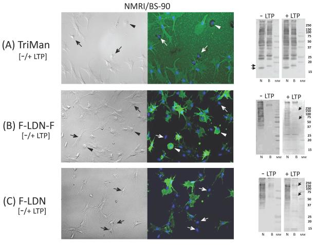
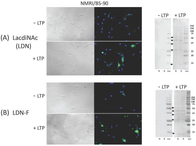
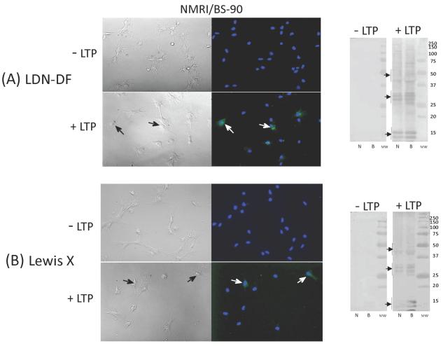
Host lectin-like recognition molecules may play an important role in innate resistance in Biomphalaria glabrata snails to larval schistosome infection, thus implicating parasite-expressed glycans as putative ligands for these lectin receptors. While host lectins may utilize specific glycan structures for parasite recognition, it also has been hypothesized that the parasite may use this system to evade immune detection by mimicking naturally-expressed host glycans, resulting in reduced immunorecognition capacity. By employing immunocytochemical (ICC) and Western blot assays using schistosome glycan-specific monoclonal antibodies (mABs) we sought to identify specific glycan epitopes (glycotopes) shared in common between larval Schistosoma mansoni and B. glabrata hemocytes, the primary immune effector cells in snails. Results confirmed the presence of selected larval glycotopes on subpopulations of hemocytes by ICC and association with numerous hemocyte proteins by Western blot analyses, including a trimannosyl core N-glycan (TriMan), and two fucosylated lacdiNAc (LDN) variants, F-LDN and F-LDN-F. Snail strain differences were seen in the prevalence of constitutively expressed F-LDN on hemocytes, and in the patterns of protein immunoreactivity with these mABs. In contrast, there was little to no hemocyte reactivity with mABs for Lewis X (LeX), LDN, LDN-F or LDN-DF. When intact hemocytes were exposed to larval transformation products (LTPs), distinct cell subpopulations displayed weak (LeX, LDN-DF) to moderate (LDN, LDN-F) glycotope reactivity by ICC, including snail strain differences in the prevalence of LDN-reactive cellular subsets. Far-Western blot analyses of the hemocytes following exposure to larval transformation proteins (LTPs) also revealed multiple mAB-reactive hemocyte protein bands for LeX, LDN, LDN-F, and LDN-DF. These results demonstrate the existence of complex patterns of shared larval glycan constitutively expressed on hemocytes and their proteins, as well as the ability of hemocytes to acquire shared glycans by the selective binding of parasite-released LTP. Unraveling the functional significance of these naturally expressed and acquired shared glycans on specific hemocyte populations represents an important challenge for future investigations.
Copyright © 2012 Elsevier Inc. All rights reserved.
Figures
Similar articles
-
Glycotope sharing between snail hemolymph and larval schistosomes: larval transformation products alter shared glycan patterns of plasma proteins.PLoS Negl Trop Dis. 2012;6(3):e1569. doi: 10.1371/journal.pntd.0001569. Epub 2012 Mar 20. PLoS Negl Trop Dis. 2012. PMID: 22448293 Free PMC article.
-
Glycotope analysis in miracidia and primary sporocysts of Schistosoma mansoni: differential expression during the miracidium-to-sporocyst transformation.Int J Parasitol. 2009 Oct;39(12):1331-44. doi: 10.1016/j.ijpara.2009.06.002. Epub 2009 Jun 21. Int J Parasitol. 2009. PMID: 19545571 Free PMC article.
-
Larval Schistosoma mansoni excretory-secretory glycoproteins (ESPs) bind to hemocytes of Biomphalaria glabrata (Gastropoda) via surface carbohydrate binding receptors.J Parasitol. 2001 Aug;87(4):786-93. doi: 10.1645/0022-3395(2001)087[0786:LSMESG]2.0.CO;2. J Parasitol. 2001. PMID: 11534642
-
Epigenetic modulation, stress and plasticity in susceptibility of the snail host, Biomphalaria glabrata, to Schistosoma mansoni infection.Int J Parasitol. 2016 Jun;46(7):389-94. doi: 10.1016/j.ijpara.2016.03.003. Epub 2016 Apr 4. Int J Parasitol. 2016. PMID: 27056272 Review.
-
Immuno-molecular profile for Biomphalaria glabrata/Schistosoma mansoni interaction.Dev Comp Immunol. 2024 Jan;150:105083. doi: 10.1016/j.dci.2023.105083. Epub 2023 Oct 17. Dev Comp Immunol. 2024. PMID: 37852455 Review.
Cited by
-
Study of surface carbohydrates in Galba truncatula tissues before and after infection with Fasciola hepatica.Mem Inst Oswaldo Cruz. 2016 Jul 4;111(7):475-83. doi: 10.1590/0074-02760160141. Mem Inst Oswaldo Cruz. 2016. PMID: 27384082 Free PMC article.
-
Mollusc N-glycosylation: Structures, Functions and Perspectives.Biomolecules. 2021 Dec 3;11(12):1820. doi: 10.3390/biom11121820. Biomolecules. 2021. PMID: 34944464 Free PMC article. Review.
-
Schistosomes and snails: a molecular encounter.Front Genet. 2014 Jul 21;5:230. doi: 10.3389/fgene.2014.00230. eCollection 2014. Front Genet. 2014. PMID: 25101114 Free PMC article. Review.
-
RNA-Seq reveals infection-induced gene expression changes in the snail intermediate host of the carcinogenic liver fluke, Opisthorchis viverrini.PLoS Negl Trop Dis. 2014 Mar 27;8(3):e2765. doi: 10.1371/journal.pntd.0002765. eCollection 2014 Mar. PLoS Negl Trop Dis. 2014. PMID: 24676090 Free PMC article.
-
Deciphering the glycogenome of schistosomes.Front Genet. 2014 Aug 5;5:262. doi: 10.3389/fgene.2014.00262. eCollection 2014. Front Genet. 2014. PMID: 25147556 Free PMC article. Review.
References
-
- Bayne CJ, Buckley PM, DeWan PC. Schistosoma mansoni: cytotoxicity of hemocytes from susceptible snail hosts for sporocysts in plasma from resistant Biomphalaria glabrata. Exp. Parasitol. 1980;50:409–146. - PubMed
-
- Cavalcanti MGS, Filho FC, Mendonca AM, Duarte GR, Barbosa CC, De Castro CM, Alves LC, Brayner FA. Morphological characterization of hemocytes from Biomphalaria glabrata and Biomphalaria straminea. Micron. 2012;43:285–291. - PubMed
-
- Cheng TC, Auld KR. Hemocytes of the pulmonate gastropod Biomphalaria glabrata. J. Invertebr. Pathol. 1977;30:119–122. - PubMed
Publication types
MeSH terms
Substances
Grants and funding
LinkOut - more resources
Full Text Sources
Other Literature Sources
Research Materials
Miscellaneous
