

Optical imaging of periostin enables early endoscopic detection and characterization of esophageal cancer in mice
- PMID: 23085486
- PMCID: PMC3624041
- DOI: 10.1053/j.gastro.2012.10.030
Optical imaging of periostin enables early endoscopic detection and characterization of esophageal cancer in mice
Abstract
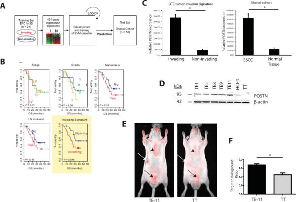
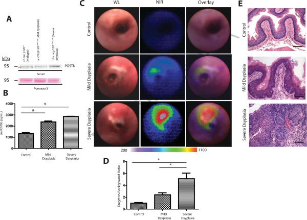
Imaging strategies that detect early stage esophageal squamous cell carcinoma (ESCC) could improve clinical outcomes, when combined with endoscopic approaches. Periostin is an integrin-binding protein that is important in the tumor microenvironment. We created a fluorescent-labeled antibody that recognizes periostin and binds specifically to ESCC xenograft tumors in mice. In L2-cre;p120ctnLoxP/LoxP mice, which develop squamous cell cancers that resemble human ESCC, we visualized the probe in preneoplastic and neoplastic esophageal lesions using near-infrared fluorescent imaging with upper-gastrointestinal endoscopy. Periostin might be a biomarker of the esophageal tumor microenvironment that can be used to detect preneoplastic lesions.
Copyright © 2013 AGA Institute. Published by Elsevier Inc. All rights reserved.
Figures
References
-
- Ferlay J, et al. Int J Cancer. 2010;127:2893–917. - PubMed
-
- Mayer P, et al. The New England Journal of Medicine. 2003:2241–52. - PubMed
-
- Wang LS, et al. Am J Gastroenterol. 1999;94:1933–40. - PubMed
-
- Lao-Sirieix P, et al. Nat Rev Clin Oncol. 2012;9:278–87. - PubMed
-
- Bird-Lieberman EL, et al. Nat Med. 2012;18:315–21. - PubMed
Publication types
MeSH terms
Grants and funding
- T32-CA115299/CA/NCI NIH HHS/United States
- P30 DK050306/DK/NIDDK NIH HHS/United States
- F32-CA162719/CA/NCI NIH HHS/United States
- P01-CA098101/CA/NCI NIH HHS/United States
- P01 CA098101/CA/NCI NIH HHS/United States
- T32-DK007066/DK/NIDDK NIH HHS/United States
- P30 CA016520/CA/NCI NIH HHS/United States
- T32 CA115299/CA/NCI NIH HHS/United States
- U01-CA14305603/CA/NCI NIH HHS/United States
- U01 CA143056/CA/NCI NIH HHS/United States
- P30-DK050306/DK/NIDDK NIH HHS/United States
- T32 DK007066/DK/NIDDK NIH HHS/United States
LinkOut - more resources
Full Text Sources
Other Literature Sources
Medical
