

doi: 10.1038/nmeth.2211.
Epub 2012 Oct 21.
Membrane-protein binding measured with solution-phase plasmonic nanocube sensors
Affiliations
- PMID: 23085614
- PMCID: PMC3703907
- DOI: 10.1038/nmeth.2211
Item in Clipboard
Membrane-protein binding measured with solution-phase plasmonic nanocube sensors
Nat Methods.
2012 Dec.
Abstract
We describe a solution-phase sensor of lipid-protein binding based on localized surface plasmon resonance (LSPR) of silver nanocubes. When silica-coated nanocubes are mixed in a suspension of lipid vesicles, supported membranes spontaneously assemble on their surfaces. Using a standard laboratory spectrophotometer, we calibrated the LSPR peak shift due to protein binding to the membrane surface and then characterized the lipid-binding specificity of a pleckstrin homology domain protein.
Conflict of interest statement
The authors declare no competing financial interests.
Figures
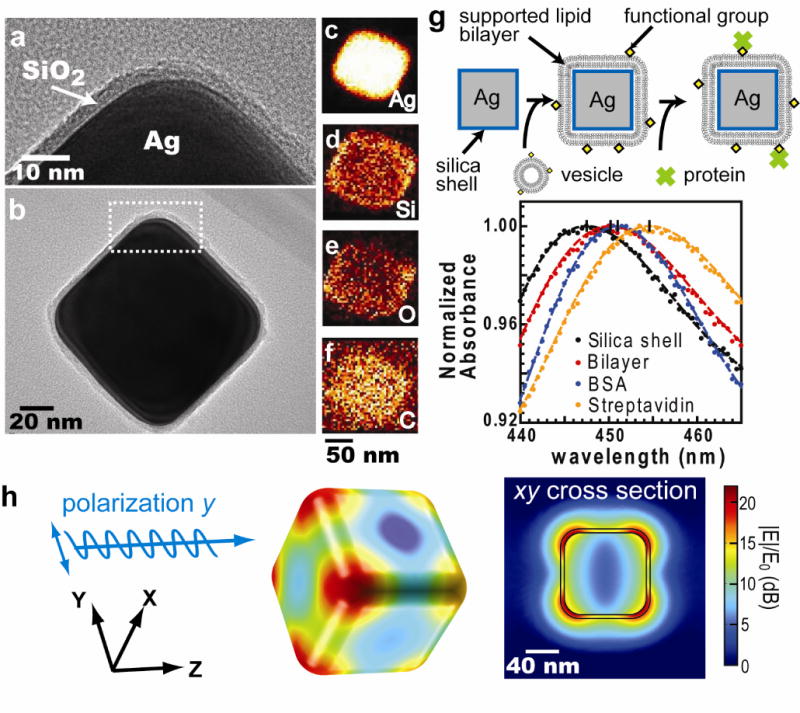
The physical properties of Ag@SiO2 core-shell nanocube. (a) & (b) TEM images of Ag@SiO2 nanocube. (a) is the close-up image of figure (b). (c)~(f) The elemental maps obtained by high-angle annular dark field scanning TEM (HAADF-STEM) with energy dispersive x-ray spectroscopy (EDS). (c) to (f) represent silver, silicon, oxygen, and carbon, respectively. (g) Top: Detection procedure of nanocube sensors. Supported lipid bilayers are formed by vesicle fusion onto the silica surface, and protein binding is monitored by shifts in the LSPR extinction spectrum. Bottom: Typical spectra of membrane coverage and protein binding to the membrane surfaces. Sequential addition of lipid vesicles, BSA, and streptavidin causes LSPR red shifts. (h) Electric field norm (|E|/E0) in decibel (dB) of a nanocube at resonance (n = 1.33303, λ0 = 474 nm) computed using finite-element analysis.
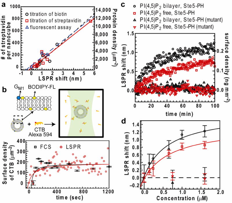
Calibration of the nanocube assay. (a) Relation between LSPR shift and number of streptavidin per nanocube (left vertical axis) and surface density (right axis) measured by titration of biotinyl-cap-PE, titration of streptavidin, and fluorescence measurement of streptavidin concentration. Linear fit slopes are reported in Supplementary Table 1. (b) Top: Concentrations of bound and unbound CTB are detected by multi-component FCS. Alexa 594-CTB binds to vesicles (average diameter 120 nm) containing 0.5% GM1 and 0.5% BODIPY-FL-DHPE lipids. BODIPY-FL-DHPE was used to determine the average number of vesicles diffusing within the excitation spot. Bottom: Binding kinetics measured by multi-component FCS and nanocube assay. (Error bar of FCS, n = 20, mean ± s.d.) CTB surface density was respectively calculated from known vesicle size and LSPR response to protein mass change in streptavidin-biotin systems (0.191 ng mm−2 nm−1). (c) Binding kinetics of wild-type and R407S K411S mutant of GST-Ste5 PH to different membrane surfaces. Concentrations of GST-Ste5 PH = 1.6 μM; GST-Ste5 PH mutant = 1.6 μM) (d) Equilibrium binding curves of GST-Ste5 PH to bilayers Kd = 0.49 ± 0.33 μM (PI(4,5)P2 bilayer) and 1.6 ± 0.45 μM (PI(4,5)P2-free bilayer) (n = 3, mean ± s.e.m.) Error limits of Kd are derived from the statistical error of curve fitting.
References
Publication types
MeSH terms
Substances
Grants and funding
LinkOut - more resources
Full Text Sources
Other Literature Sources
