Review
. 2012 Dec;23(9):946-53.
doi: 10.1016/j.semcdb.2012.10.001. Epub 2012 Oct 17.

Epithelial stem cells and implications for wound repair

Affiliations
Review

Epithelial stem cells and implications for wound repair

Maksim V Plikus et al. Semin Cell Dev Biol. 2012 Dec.

Abstract

Activation of epithelial stem cells and efficient recruitment of their proliferating progeny plays a critical role in cutaneous wound healing. The reepithelialized wound epidermis has a mosaic composition consisting of progeny that can be traced back both to epidermal and several types of hair follicle stem cells. The contribution of hair follicle stem cells to wound epidermis is particularly intriguing as it involves lineage identity change from follicular to epidermal. Studies from our laboratory show that hair follicle-fated bulge stem cells commit only transient amplifying epidermal progeny that participate in the initial wound re-epithelialization, but eventually are outcompeted by other epidermal clones and largely disappear after a few months. Conversely, recently described stem cell populations residing in the isthmus portion of hair follicle contribute long-lasting progeny toward wound epidermis and, arguably, give rise to new interfollicular epidermal stem cells. The role of epithelial stem cells during wound healing is not limited to regenerating stratified epidermis. By studying regenerative response in large cutaneous wounds, our laboratory uncovered that epithelial cells in the center of the wound can acquire greater morphogenetic plasticity and, together with the underlying wound dermis, can engage in an embryonic-like process of hair follicle neogenesis. Future studies should uncover the cellular and signaling basis of this remarkable adult wound regeneration phenomenon.


Figures

Figure 1
Figure 1. Heterogeneity of epidermal and hair follicle stem cells
Skin epithelia feature distinct stem cell populations both in epidermis and, most prominently, in hair follicles. Epithelial stem cells in different micro-anatomical locations have different lineage potentials. Stem cells in the follicular infundibulum and inter-follicular epidermis physiologically are restricted to epidermal fate. Inter-follicular epidermal stem cells can be identified as slow-cycling in label retention studies, but distinct markers remain elusive. The isthmus and junctional zone of hair follicles harbor several distinct epithelial cell populations. Most prominent among them are Lrig1+ (yellow), Gli1+ and Lgr6+ stem cells (green), all of which physiologically maintain the isthmus and contribute to sebaceous gland, infundibulum and in some instance to inter-follicular epidermis. Blimp1 identifies unipotent sebaceous gland progenitors (orange). The bulge stem cells (blue) normally contribute to all hair follicle lineages and can be identified based on the expression of Krt15, CD200, Lgr5, CD34, Sox9, Lhx2, Tcf3 and Nfatc1. The secondary germ of telogen hair follicles (purple) contains committed hair follicle-fated progenitors that express CD200, Gli1 and Lgr5.
Figure 2
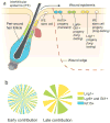
Figure 2. Contribution of hair follicle stem cells to wound epidermis
a) Upon wounding, both Lrig1+ (yellow) and Lgr6+ (green) stem cells and Krt15+bulge stem cells (blue) within hair follicles along the wound edge become activated and generate progeny that migrate out of the follicles and participate in rapid wound re-epithelialization. b) While bulge stem cells contribute only transiently to amplifying epidermal progeny, Lrig1+, Lgr6+ stem cells are able to generate long-lasting epidermal clones. As the consequence of these dynamics, bulge stem cell progeny in wound epidermis significantly decline over time.
Figure 3
Figure 3. The role of epithelial stem cells in wounding-induced hair follicle neogenesis
Following re-epithelialization of large full-thickness wounds, new hair follicles develop from the basal cells of the wound epidermis via a process of embryonic-like neogenesis. Progeny of the Krt15+ bulge stem cells from the peri-wound hair follicles do not participate in making de novo hair follicles, consistent with their transient involvement in wound re-epithelialization. While currently the original lineage identity of the epithelial stem cells that contribute to hair follicle neogenesis is unknown, preliminary data points at the involvement of isthmus stem cells or possibly interfollicular stem cells.

References

    1. Ansell DM, Kloepper JE, Thomason HA, Paus R, Hardman MJ. Exploring the “Hair Growth-Wound Healing Connection”: Anagen Phase Promotes Wound Re-Epithelialization. J Investig Dermatol. 2011;131:518–28. - PubMed
    1. Borue X, Lee S, Grove J, Herzog EL, Harris R, Diflo T, Glusac E, Hyman K, Theise ND, Krause DS. Bone marrow-derived cells contribute to epithelial engraftment during wound healing. Am J Pathol. 2004;165:1767–72. - PMC - PubMed
    1. Brittan M, Braun KM, Reynolds LE, Conti FJ, Reynolds AR, Poulsom R, Alison MR, Wright NA, Hodivala-Dilke KM. Bone marrow cells engraft within the epidermis and proliferate in vivo with no evidence of cell fusion. J Pathol. 2005;205:1–13. - PubMed
    1. Brownell I, Guevara E, Bai CB, Loomis CA, Joyner AL. Nerve-derived sonic hedgehog defines a niche for hair follicle stem cells capable of becoming epidermal stem cells. Cell Stem Cell. 2011;8:552–65. - PMC - PubMed
    1. Cotsarelis G, Sun TT, Lavker RM. Label-retaining cells reside in the bulge area of pilosebaceous unit: implications for follicular stem cells, hair cycle, and skin carcinogenesis. Cell. 1990;61:1329– 37. - PubMed

MeSH terms