

Anesthetics interfere with the polarization of developing cortical neurons
- PMID: 23085784
- PMCID: PMC3479440
- DOI: 10.1097/ANA.0b013e31826a03a6
Anesthetics interfere with the polarization of developing cortical neurons
Abstract
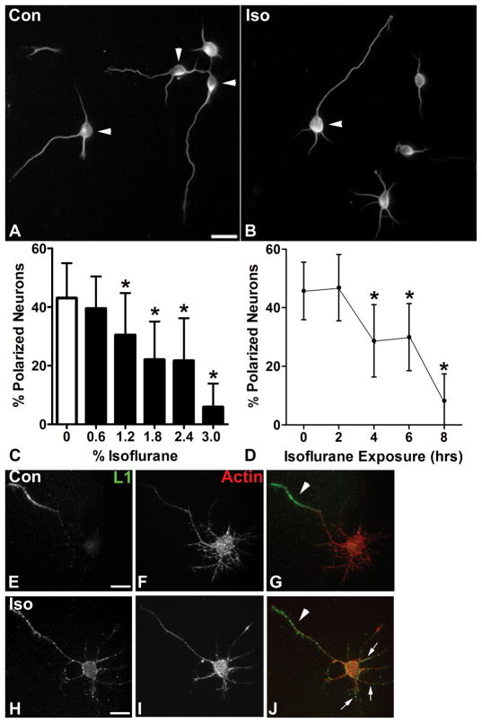
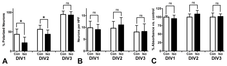
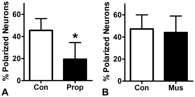
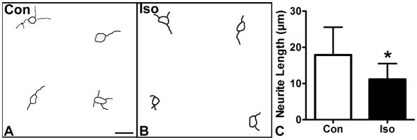
Numerous studies from the clinical and preclinical literature indicate that general anesthetic agents have toxic effects on the developing brain, but the mechanism of this toxicity is still unknown. Previous studies have focused on the effects of anesthetics on cell survival, dendrite elaboration, and synapse formation, but little attention has been paid to possible effects of anesthetics on the developing axon. Using dissociated mouse cortical neurons in culture, we found that isoflurane delays the acquisition of neuronal polarity by interfering with axon specification. The magnitude of this effect is dependent on isoflurane concentration and exposure time over clinically relevant ranges, and it is neither a precursor to nor the result of neuronal cell death. Propofol also seems to interfere with the acquisition of neuronal polarity, but the mechanism does not require activity at GABAA receptors. Rather, the delay in axon specification likely results from a slowing of the extension of prepolarized neurites. The effect is not unique to isoflurane as propofol also seems to interfere with the acquisition of neuronal polarity. These findings demonstrate that anesthetics may interfere with brain development through effects on axon growth and specification, thus introducing a new potential target in the search for mechanisms of pediatric anesthetic neurotoxicity.
Conflict of interest statement
Conflict of Interest: None
Figures
Similar articles
-
Anesthetics interfere with axon guidance in developing mouse neocortical neurons in vitro via a γ-aminobutyric acid type A receptor mechanism.Anesthesiology. 2013 Apr;118(4):825-33. doi: 10.1097/ALN.0b013e318287b850. Anesthesiology. 2013. PMID: 23364597 Free PMC article.
-
Isoflurane impairs the capacity of astrocytes to support neuronal development in a mouse dissociated coculture model.J Neurosurg Anesthesiol. 2014 Oct;26(4):363-8. doi: 10.1097/ANA.0000000000000119. J Neurosurg Anesthesiol. 2014. PMID: 25191957 Free PMC article.
-
Comparison of neurodegeneration and cognitive impairment in neonatal mice exposed to propofol or isoflurane.PLoS One. 2014 Jun 16;9(6):e99171. doi: 10.1371/journal.pone.0099171. eCollection 2014. PLoS One. 2014. PMID: 24932894 Free PMC article.
-
Actions of Propofol on Neurons in the Cerebral Cortex.J Nippon Med Sch. 2017;84(4):165-169. doi: 10.1272/jnms.84.165. J Nippon Med Sch. 2017. PMID: 28978896 Review.
-
Strategies and experimental models for evaluating anesthetics: effects on the developing nervous system.Anesth Analg. 2008 Jun;106(6):1643-58. doi: 10.1213/ane.ob013e3181732c01. Anesth Analg. 2008. PMID: 18499593 Review.
Cited by
-
Pilose antler polypeptide protects against sevoflurane‑mediated neurocyte injury.Mol Med Rep. 2018 Dec;18(6):5353-5360. doi: 10.3892/mmr.2018.9582. Epub 2018 Oct 24. Mol Med Rep. 2018. PMID: 30365108 Free PMC article.
-
An allosteric propofol-binding site in kinesin disrupts kinesin-mediated processive movement on microtubules.J Biol Chem. 2018 Jul 20;293(29):11283-11295. doi: 10.1074/jbc.RA118.002182. Epub 2018 May 29. J Biol Chem. 2018. PMID: 29844014 Free PMC article.
-
Sevoflurane exposure during the second trimester induces neurotoxicity in offspring rats by hyperactivation of PARP-1.Psychopharmacology (Berl). 2022 Sep;239(9):3031-3045. doi: 10.1007/s00213-022-06188-4. Epub 2022 Jul 20. Psychopharmacology (Berl). 2022. PMID: 35859039
-
The neuroprotective effects of remifentanil on isoflurane-induced apoptosis in the neonatal rat brain.Am J Transl Res. 2017 Oct 15;9(10):4521-4533. eCollection 2017. Am J Transl Res. 2017. PMID: 29118914 Free PMC article.
-
Early postnatal exposure to isoflurane causes cognitive deficits and disrupts development of newborn hippocampal neurons via activation of the mTOR pathway.PLoS Biol. 2017 Jul 6;15(7):e2001246. doi: 10.1371/journal.pbio.2001246. eCollection 2017 Jul. PLoS Biol. 2017. PMID: 28683067 Free PMC article.
References
-
- Levin ED, Uemura E, Bowman RE. Neurobehavioral toxicology of halothane in rats. Neurotoxicol Teratol. 1991;13:461–70. - PubMed
-
- Quimby KL, Aschkenase LJ, Bowman RE, et al. Enduring learning deficits and cerebral synaptic malformation from exposure to 10 parts of halothane per million. Science. 1974;185:625–7. - PubMed
-
- Uemura E, Ireland WP, Levin ED, et al. Effects of halothane on the development of rat brain: a golgi study of dendritic growth. Exp Neurol. 1985;89:503–19. - PubMed
Publication types
MeSH terms
Substances
Grants and funding
LinkOut - more resources
Full Text Sources
