. 2013 Jan 1;3(1):a006569.
doi: 10.1101/cshperspect.a006569.

VEGF and Notch in tip and stalk cell selection

Affiliations

VEGF and Notch in tip and stalk cell selection

Raquel Blanco et al. Cold Spring Harb Perspect Med. .

Abstract

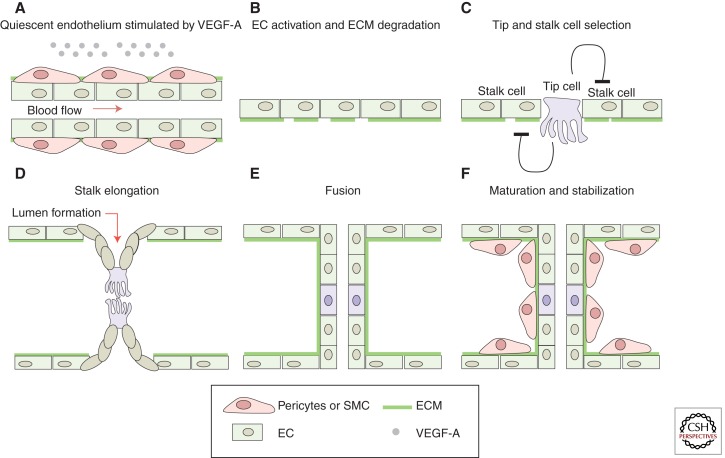
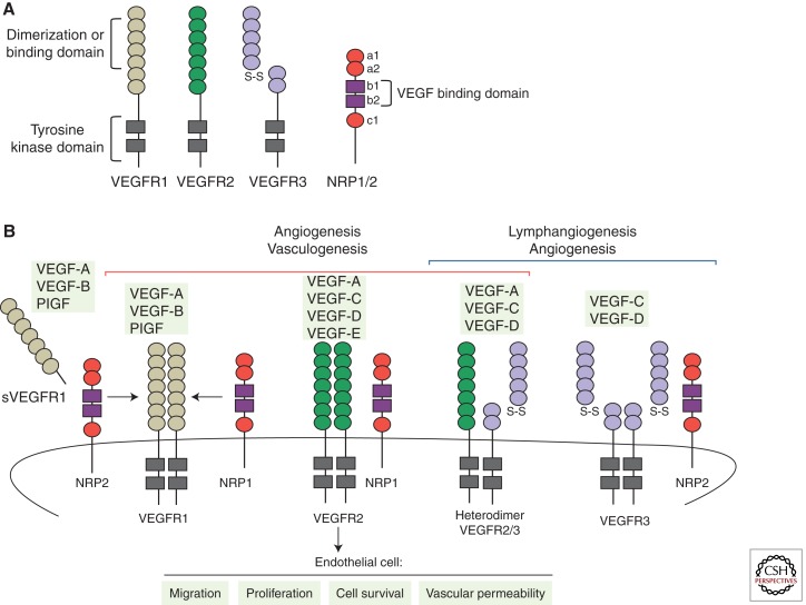
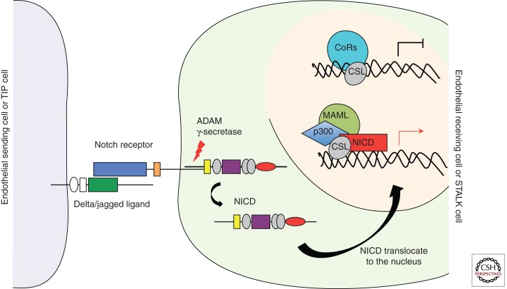
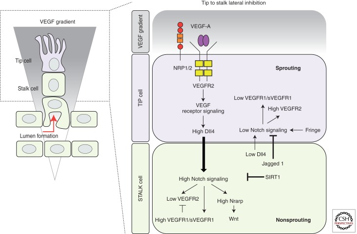
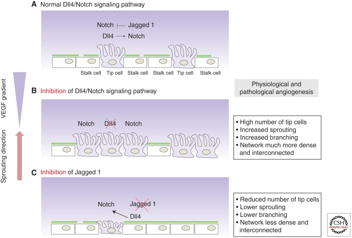
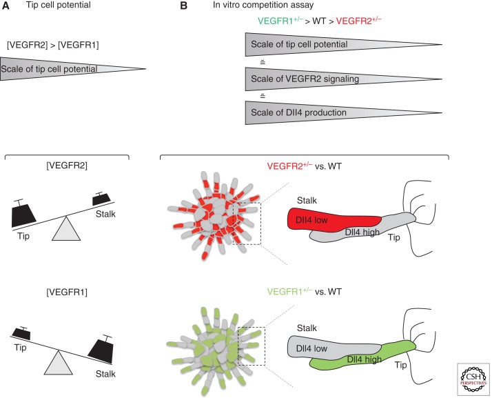
Sprouting angiogenesis is a dynamic process in which endothelial cells collectively migrate, shape new lumenized tubes, make new connections, and remodel the nascent network into a hierarchically branched and functionally perfused vascular bed. Endothelial cells in the nascent sprout adopt two distinct cellular phenotypes--known as tip and stalk cells--with specialized functions and gene expression patterns. VEGF and Notch signaling engage in an intricate cross talk to balance tip and stalk cell formation and to regulate directed tip cell migration and stalk cell proliferation. In this article, we summarize the current knowledge and implications of the tip/stalk cell concepts and the quantitative and dynamic integration of VEGF and Notch signaling in tip and stalk cell selection.


Figures

Figure 1.
Figure 1.
Schematic model of sprout initiation, vessel branching, and maturation. Angiogenesis is activated in response to local tissue hypoxia. The hypoxic tissue releases endothelial growth factor, that is, VEGF-A, which (re)activates the quiescent endothelial cells (ECs). (A) At the cellular level, the angiogenic initiation requires the degradation of the extra cellular matrix (ECM) (B) as well as the specification of the (re)activated ECs into tip and stalk cells (C). ECs proliferate and collectively invade the hypoxic tissue while they remain connected to the original vascular network. (D) In the nascent sprout, the tip cells, characterized by their migratory behavior and dynamic filopodia, lead the sprout toward the VEGF-A source, whereas the stalk cells proliferate to support sprout elongation. The tip cells connect the new sprouts into a functional vessel loop. (E) The new connection between different sprouts occurs through tip cell fusion (anastomosis). Formation of the vascular lumen initiates blood flow, increases tissue oxygenation, and, in turn, reduces the release of endothelial growth factors, supporting the establishment of quiescence. (F) Vessel maturation and stabilization proceed with the recruitment of mural cells (pericytes) and the deposition of ECM.
Figure 2.
Figure 2.
VEGFs, VEGF receptors, and coreceptors. (A) Outline of the structural domains of the different VEGFRs and coreceptors Nrp1 and Nrp2. (B) Schematic representation of the binding specificity of the different VEGF family members (VEGF-A, -B, -C, -D, -E, and PlGF) to the tyrosine kinase receptors, VEGFR1, VEGFR2, and VEGFR3. Interactions between VEGFRs and the coreceptors Nrp1 or Nrp2 are also shown in the figure. Activation of VEGFR1 and VEGFR2 regulates vasculogenesis and angiogenesis. However, activation of VEGFR3 stimulates lymphangiogenesis and embryonic angiogenesis. In particular, during sprouting angiogenesis VEGFR2 is the principal mediator of VEGF-A signaling, activating a variety of downstream signaling pathways that regulate endothelial cell migration, survival, proliferation, and tube formation. In contrast, VEGFR1 or sVEGFR1, acting as a decoy receptor, limits the VEGF activity in the vascular endothelium.
Figure 3.
Figure 3.
Canonical Notch signaling pathway. The Notch pathway is an evolutionarily conserved intercellular signaling mechanism implicated in cell fate determination and differentiation of endothelial cells. Notch signaling begins with receptor–ligand interaction between neighboring cells. This interaction triggers a series of proteolytic cleavages of the Notch receptor. The final one, catalyzed by the γ-secretase complex, releases the active Notch intercellular domain (NICD) from the cell membrane. NICD is translocated to the nucleus, where it interacts directly with the transcription factor CSL. Following binding of NICD to CSL, the repressor complex is converted into an activating complex by displacing corepressors and recruiting coactivators. After the coactivator complex is recruited, the transcription of promoters that contains CSL-binding elements is induced. Hairy/enhancer of split (HES) and HES-related proteins (HEY/HRT/HERP) family genes are included among the target genes of Notch signaling. Dll4 ligand is also a target gene regulated by the Notch signaling pathway.
Figure 4.
Figure 4.
Tip/stalk cell specification during sprouting angiogenesis. During sprouting angiogenesis, VEGF and Notch signaling pathways are implicated in the specification of tip and stalk cells in the vascular endothelium. VEGF interacts with VEGFR2, expressed at the surface of the endothelial cells of the quiescent vessels. Nrp1 modulates the VEGF signaling output, enhancing the binding activity and signaling of VEGF through VEGFR2. Under VEGF stimulation, Dll4 expression is up-regulated in the tip cells. In turn, Dll4 ligand activates Notch signaling in the stalk, consequently suppressing the tip cell phenotype. Notch signaling activation reduces VEGFR2 expression and increases VEGFR1/sVEGFR1 levels as well as the expression of different Notch target genes (e.g., Notch-regulated ankyrin repeat protein [Nrarp]). In contrast, the tip cell receives low Notch signaling, allowing high expression of VEGFR2 and Nrp1, but low VEGFR1. Contrary to Dll4, Jagged1 ligand is expressed by the stalk cells. Jagged1 antagonizes Dll4–Notch signaling in the sprouting front when the Notch receptor is modified by the glycosyltranferase Fringe, thereby enhancing differential Notch activity between tip and stalk cells. The duration and amplitude of the Notch signal are modulated by the histone deacetylase SIRT1, which, by direct deacetylation, primes the Notch intracellular domain for ubiquitination and degradation.
Figure 5.
Figure 5.
Dll4 and Jagged1 show antagonistic function during sprouting angiogenesis. Tip/stalk cell selection is mediated via Dll4/Notch lateral inhibition between the ECs that constitute the vasculature, resulting in the typical salt-and-pepper distribution of tip cells along the vascular endothelium. (A) For a detailed explanation about Dll4/Notch lateral inhibition see Figure 4. (B) Genetic or pharmacological inhibition of Dll4 deregulates the tip/stalk selection process; dramatically increases the number of tip cells, the number of sprouts and branching, resulting in a hyperdense and interconnected plexus. (C) The opposite observation has been made after Jagged1 inhibition in physiological and pathological angiogenesis, as Jagged1 antagonizes reciprocal Dll4-mediated Notch activation from the stalk to the tip cell.
Figure 6.
Figure 6.
VEGFR1 and VEGFR2 levels regulate the tip cell potential in the vascular endothelium. (A) Schematic representation of how VEGFRs levels affect tip/stalk cell potential. (B) Outline of the genetic mosaic-sprouting assay in vitro between wild-type (WT) and VEGFR2 or VEGFR1 heterozygous endothelial cells. Cells with higher VEGFR2 levels or lower VEGFR1 levels preferentially acquire the lead position (tip cell) in the sprout. VEGFR levels modulate the expression of Dll4 ligand during the selection process. VEGFR2+/− cells produce less Dll4 mRNA and protein when competing with wild-type cells, which reduces their ability to compete for the tip cell phenotype. The opposite is true for VEGFR1+/− cells in competition with wild-type cells.

Similar articles

Cited by

References

    1. Autiero M, Waltenberger J, Communi D, Kranz A, Moons L, Lambrechts D, Kroll J, Plaisance S, De Mol M, Bono F, et al. 2003. Role of PlGF in the intra- and intermolecular cross talk between the VEGF receptors Flt1 and Flk1. Nat Med 9: 936–943 - PubMed
    1. Barleon B, Sozzani S, Zhou D, Weich HA, Mantovani A, Marme D 1996. Migration of human monocytes in response to vascular endothelial growth factor (VEGF) is mediated via the VEGF receptor flt-1. Blood 87: 3336–3343 - PubMed
    1. Bellon A, Luchino J, Haigh K, Rougon G, Haigh J, Chauvet S, Mann F 2010. VEGFR2 (KDR/Flk1) signalling mediates axon growth in response to semaphorin 3E in the developing brain. Neuron 66: 205–219 - PubMed
    1. Benedito R, Roca C, Sorensen I, Adams S, Gossler A, Fruttiger M, Adams RH 2009. The notch ligands Dll4 and Jagged1 have opposing effects on angiogenesis. Cell 137: 1124–1135 - PubMed
    1. Bentley K, Gerhardt H, Bates PA 2008. Agent-based simulation of notch-mediated tip cell selection in angiogenic sprout initialisation. J Theor Biol 250: 25–36 - PubMed

Publication types

MeSH terms

Substances

LinkOut - more resources