Review
. 2013 Jun;32(1-2):5-24.
doi: 10.1007/s10555-012-9407-3.

What underlies the diversity of brain tumors?

Affiliations
Review

What underlies the diversity of brain tumors?

Fredrik J Swartling et al. Cancer Metastasis Rev. 2013 Jun.

Abstract

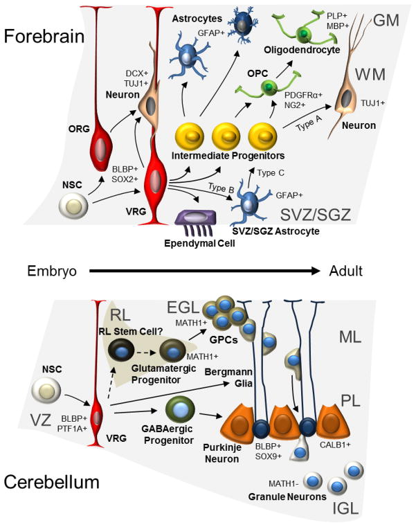
Glioma and medulloblastoma represent the most commonly occurring malignant brain tumors in adults and in children, respectively. Recent genomic and transcriptional approaches present a complex group of diseases and delineate a number of molecular subgroups within tumors that share a common histopathology. Differences in cells of origin, regional niches, developmental timing, and genetic events all contribute to this heterogeneity. In an attempt to recapitulate the diversity of brain tumors, an increasing array of genetically engineered mouse models (GEMMs) has been developed. These models often utilize promoters and genetic drivers from normal brain development and can provide insight into specific cells from which these tumors originate. GEMMs show promise in both developmental biology and developmental therapeutics. This review describes numerous murine brain tumor models in the context of normal brain development and the potential for these animals to impact brain tumor research.


Figures

Figure 1
Figure 1. Cells involved in normal brain development from embryo to adult
The schematic figure shows regions and cells of origin for glioma (in forebrain, above) and MB (in cerebellum, below). Processes of the radial glia (RG) serve as guides for migrating more immature neurons [196,197]. RG born from neuroepithelial stem cells could give rise to basically all other cell types including ependymal cells that line the ventricles. However, they first give rise to more restricted intermediate progenitors that determine the path for astrocytes (protoplasmic (in GM) or fibrous (in WM)), oligodendrocytes and neurons. Radial glia cells form neural stem cells, specifically called type B cells (SVZ astrocytes) that reside in the subventricular zone (SVZ) even in the adult forebrain [12]. Similar adult neural stem cells also exist in the dentate gyrus granule cell layer in the hippocampus [13]. Radial glia form neural stem cells that can give rise to multiple cerebellar cell types, and also more directly (around birth) convert into Bergmann glia that keep their extended processes even in adult brain. In cerebellum, granule precursor cells are also formed and divide in the external germinal layer (EGL) from the boost of SHH-producing large Purkinje neurons. Before the cerebellar EGL layer disappears (around three weeks of age in mice) the GPCs that constitute EGL migrate down the Bergmann glial processes and pass Purkinje neurons before they settle down as more differentiated granule neurons in the IGL. GM: Grey matter; WM: White matter; NSC: Neuroepithelial stem cell; ORG: Outer SVZ radial glia-like cells; VRG: ventricular epithelium radial glia; OPC: Oligodendrocyte precursor cell; SVZ: Subventricular zone; SGZ: Subgranular zone; VZ; Ventricular zone at 4th ventricle; RL: Rhombic lip; EGL: External germinal layer; GPC: Granule cell progenitor; ML: Molecular layer; PL: Purkinje cell layer; IGL: Internal granular layer. Examples of markers that are expressed, or not expressed, in the various cell types are shown followed by + or −, respectively.
Figure 2
Figure 2. Common molecular pathways of glioma
Major molecular pathways altered in glioma, starting with receptor tyrosine kinases (RTKs) on the cell surface, cytosolic proteins and nuclear proteins/transcription factors. Shown are also the relationship between these brain cancer proteins and their mechanisms of action. Proteins surrounded by broken lines identify glioma pathways that are not yet modeled in mice or described in this review.
Figure 3
Figure 3. Molecular pathways in four MB subgroups
The four major molecular pathways altered in MB. Shown are two simplified versions of the SHH and the WNT pathway that represent two of the MB subgroups. MB of Group 3 and Group 4 present some degree of MYC and MYCN alterations, respectively. Group 3 and Group 4 further present a profile of not yet identified Photoreceptor/GABAergic and Neuronal/Glutamatergic pathways, respectively. Proteins surrounded by broken lines identify MB pathways that are not yet modeled in mice or described in this review. ?: unknown pathway/mechanism.
Figure 4
Figure 4. Transgenic brain tumor models that recapitulate human brain tumors
Three examples of models where different types of brain tumors are generated from specific promoters that targets a distinct population of cells in the brain with three clinically relevant cancer genes, v-erbB (activated EGFR), PDGFB (that activates PDGFRα), Trp53 as well as MYCN. A) Tumor cells from an S100β-v-erbB-driven oligodendroglioma with typical round nuclei and perinuclear cytoplasmic retraction (black arrows). [61] B) A GFAP-PDGFB-driven Trp53 null GBM with characteristic pseudopalisadation (black arrowheads) surrounding a necrotic area (N) [76]. C) A Glt1-MYCN-driven LC/A MB with atypical large cells (white arrow) [147].
Figure 5
Figure 5. New animal models for human brain tumors
Useful features for great brain tumor models and models that are still missing or ongoing.

References

    1. Lumsden A, Krumlauf R. Patterning the vertebrate neuraxis. Science. 1996;274(5290):1109–1115. - PubMed
    1. Liu A, Niswander LA. Bone morphogenetic protein signalling and vertebrate nervous system development. Nat Rev Neurosci. 2005;6(12):945–954. - PubMed
    1. Rowitch DH, Kriegstein AR. Developmental genetics of vertebrate glial-cell specification. Nature. 2010;468(7321):214–222. - PubMed
    1. Malatesta P, Hartfuss E, Gotz M. Isolation of radial glial cells by fluorescent-activated cell sorting reveals a neuronal lineage. Development. 2000;127(24):5253–5263. - PubMed
    1. Hansen DV, Lui JH, Parker PR, Kriegstein AR. Neurogenic radial glia in the outer subventricular zone of human neocortex. Nature. 2010;464(7288):554–561. - PubMed

Publication types