

Scattering anisotropy-weighted mesoscopic imaging
- PMID: 23085898
- PMCID: PMC3434765
- DOI: 10.1117/1.JBO.17.9.090501
Scattering anisotropy-weighted mesoscopic imaging
Abstract
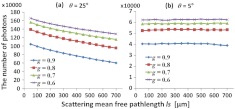
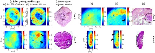
We report that when tissue images are formed via a small solid angle in the backward direction (i.e., back-directional gating), the image intensity is dominantly determined by tissue scattering anisotropy. Thus, this configuration allows for scattering anisotropy-weighted imaging that can provide an intrinsic contrast by capturing tissue structures and organizations. To demonstrate the immediate feasibility, we apply scattering anisotropy-weighted imaging to tissue blocks including basal-cell carcinomas as a pilot study. The main feature of our imaging approach is the high sensitivity to tumor locations and the simplicity for large-area visualization. We further envision that scattering anisotropy-weighted imaging could potentially be used to visualize tissue microenvironments in a mesoscopic (between microscopic and macroscopic) imaging setting.
Figures
Similar articles
-
Noninvasive measurement of scattering anisotropy in turbid materials by nonnormal incident illumination.Opt Lett. 2006 Apr 1;31(7):936-8. doi: 10.1364/ol.31.000936. Opt Lett. 2006. PMID: 16599217
-
Combined optical intensity and polarization methodology for analyte concentration determination in simulated optically clear and turbid biological media.J Biomed Opt. 2008 Jul-Aug;13(4):044037. doi: 10.1117/1.2968198. J Biomed Opt. 2008. PMID: 19021364
-
Measuring the scattering coefficient of turbid media from two-photon microscopy.Opt Express. 2013 Oct 21;21(21):25221-35. doi: 10.1364/OE.21.025221. Opt Express. 2013. PMID: 24150363
-
Comparison of simplified Monte Carlo simulation and diffusion approximation for the fluorescence signal from phantoms with typical mouse tissue optical properties.Appl Opt. 2007 Apr 1;46(10):1686-92. doi: 10.1364/ao.46.001686. Appl Opt. 2007. PMID: 17356611
-
Microscopic imaging and spectroscopy with scattered light.Annu Rev Biomed Eng. 2010 Aug 15;12:285-314. doi: 10.1146/annurev-bioeng-061008-124811. Annu Rev Biomed Eng. 2010. PMID: 20617940 Free PMC article. Review.
Cited by
-
Data-driven imaging of tissue inflammation using RGB-based hyperspectral reconstruction toward personal monitoring of dermatologic health.Biomed Opt Express. 2017 Oct 26;8(11):5282-5296. doi: 10.1364/BOE.8.005282. eCollection 2017 Nov 1. Biomed Opt Express. 2017. PMID: 29188120 Free PMC article.
References
-
- Xu Z., et al. , “Back-directional gated spectroscopic imaging for diffuse light suppression in high anisotropic media and its preclinical applications for microvascular imaging,” IEEE J. Sel. Topics Quantum Electron. 16(4), 815–823 (2010).IJSQEN10.1109/JSTQE.2009.2037160 - DOI
