

Quantitative analysis of angle-resolved scattering properties of ovarian tissue using optical coherence tomography
- PMID: 23085900
- PMCID: PMC3434469
- DOI: 10.1117/1.JBO.17.9.090503
Quantitative analysis of angle-resolved scattering properties of ovarian tissue using optical coherence tomography
Abstract
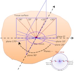
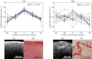
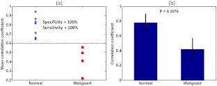
Angle-resolved optical scattering properties of ovarian tissue, on different optical coherence tomography (OCT) imaging planes, were quantitatively measured by fitting the compounded OCT A-lines into a single scattering model. Higher cross correlation value of angle-resolved scattering coefficients between different OCT imaging planes was found in normal ovaries than was present in malignant ovaries. The mean cross correlation coefficient (MCC) was introduced in this pilot study to characterize and differentiate normal, n=6, and malignant, n=4, ovaries. A specificity of 100 percent and a sensitivity of 100 percent were achieved by setting MCC threshold at 0.6. Collagen properties, within the OCT imaging penetration depth, were also qualitatively studied in terms of their content, structure and directivity. The homogeneous three-dimensional collagen fiber network, observed in the normal ovary, effectively explains the stronger cross correlation of angle-resolved scattering properties on different imaging planes while the heterogeneity, observed in the malignant ovary, suggests a weaker correlation.
Figures
Similar articles
-
An overview of optical coherence tomography for ovarian tissue imaging and characterization.Wiley Interdiscip Rev Nanomed Nanobiotechnol. 2015 Jan-Feb;7(1):1-16. doi: 10.1002/wnan.1306. Epub 2014 Oct 20. Wiley Interdiscip Rev Nanomed Nanobiotechnol. 2015. PMID: 25329515 Free PMC article. Review.
-
Optical scattering coefficient estimated by optical coherence tomography correlates with collagen content in ovarian tissue.J Biomed Opt. 2011 Sep;16(9):090504. doi: 10.1117/1.3625247. J Biomed Opt. 2011. PMID: 21950907 Free PMC article.
-
Histogram analysis of en face scattering coefficient map predicts malignancy in human ovarian tissue.J Biophotonics. 2019 Nov;12(11):e201900115. doi: 10.1002/jbio.201900115. Epub 2019 Aug 5. J Biophotonics. 2019. PMID: 31304678 Free PMC article.
-
Performance of single-scattering model versus multiple-scattering model in the determination of optical properties of biological tissue with optical coherence tomography.Appl Opt. 2010 Jun 20;49(18):3538-44. doi: 10.1364/AO.49.003538. Appl Opt. 2010. PMID: 20563206
-
Dependent and multiple scattering in transmission and backscattering optical coherence tomography.Opt Express. 2013 Dec 2;21(24):29145-56. doi: 10.1364/OE.21.029145. Opt Express. 2013. PMID: 24514466
Cited by
-
Continuous noninvasive monitoring of changes in human skin optical properties during oral intake of different sugars with optical coherence tomography.Biomed Opt Express. 2014 Feb 28;5(4):990-9. doi: 10.1364/BOE.5.000990. eCollection 2014 Apr 1. Biomed Opt Express. 2014. PMID: 24761283 Free PMC article.
-
Accuracy and safety verification of ovarian reserve assessment technique for ovarian tissue transplantation using optical coherence tomography in mice ovary.Sci Rep. 2017 Mar 8;7:43550. doi: 10.1038/srep43550. Sci Rep. 2017. PMID: 28272468 Free PMC article.
-
Beyond backscattering: optical neuroimaging by BRAD.Biomed Opt Express. 2018 May 1;9(6):2476-2494. doi: 10.1364/BOE.9.002476. eCollection 2018 Jun 1. Biomed Opt Express. 2018. PMID: 30258667 Free PMC article.
-
Preliminary human application of optical coherence tomography for quantification and localization of primordial follicles aimed at effective ovarian tissue transplantation.J Assist Reprod Genet. 2018 Apr;35(4):627-636. doi: 10.1007/s10815-018-1166-9. Epub 2018 Apr 2. J Assist Reprod Genet. 2018. PMID: 29607457 Free PMC article.
-
An overview of optical coherence tomography for ovarian tissue imaging and characterization.Wiley Interdiscip Rev Nanomed Nanobiotechnol. 2015 Jan-Feb;7(1):1-16. doi: 10.1002/wnan.1306. Epub 2014 Oct 20. Wiley Interdiscip Rev Nanomed Nanobiotechnol. 2015. PMID: 25329515 Free PMC article. Review.
References
-
- Brewer M. A., et al. , “Imaging of the ovary,” Technol. Cancer Res. Treat. 3(6), 617–627 (2004).TCRTBS - PubMed
Publication types
MeSH terms
Grants and funding
LinkOut - more resources
Full Text Sources
Medical
