. 2012 Nov;9(6):464-72.
doi: 10.1038/cmi.2012.47. Epub 2012 Oct 22.

Induction of M2-like macrophages in recipient NOD-scid mice by allogeneic donor CD4(+)CD25(+) regulatory T cells

Affiliations

Induction of M2-like macrophages in recipient NOD-scid mice by allogeneic donor CD4(+)CD25(+) regulatory T cells

Xuelian Hu et al. Cell Mol Immunol. 2012 Nov.

Abstract

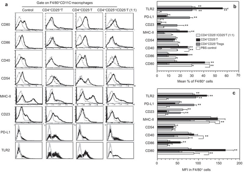
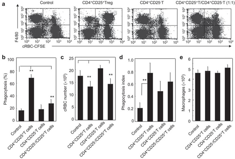
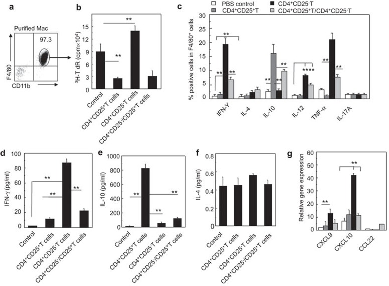
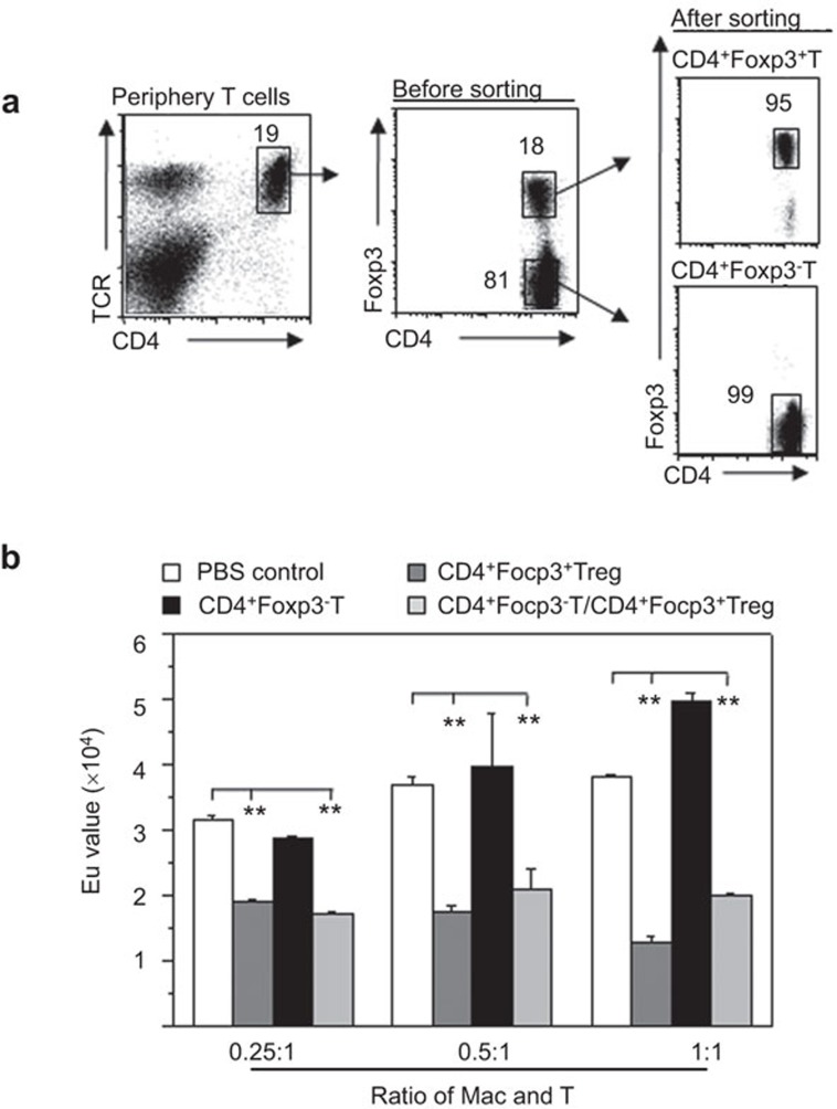
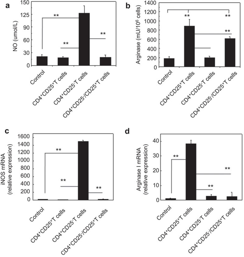
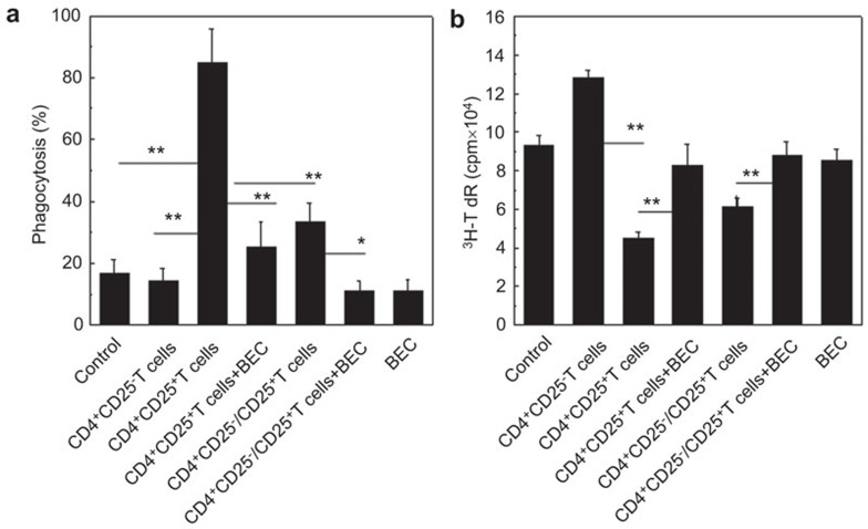
CD4(+)CD25(+) regulatory T cells (Tregs) play an important role in maintaining host immune tolerance via regulation of the phenotype and function of the innate and adaptive immune cells. Whether allogeneic CD4(+)CD25(+) Tregs can regulate recipient mouse macrophages is unknown. The effect of allogeneic donor CD4(+)CD25(+) Tregs on recipient mouse resident F4/80(+)macrophages was investigated using a mouse model in which allogeneic donor CD4(+)CD25(+) Tregs were adoptively transferred into the peritoneal cavity of host NOD-scid mice. The phenotype and function of the recipient macrophages were then assayed. The peritoneal F4/80(+) macrophages in the recipient mice that received the allogeneic CD4(+)CD25(+) Tregs expressed significantly higher levels of CD23 and programmed cell death-ligand 1(PD-L1) and lower levels of CD80, CD86, CD40 and MHC II molecules compared to the mice that received either allogeneic CD4(+)CD25(-) T cells (Teffs) or no cells. The resident F4/80(+) macrophages of the recipient mice injected with the allogeneic donor CD4(+)CD25(+) Tregs displayed significantly increased phagocytosis of chicken red blood cells (cRBCs) and arginase activity together with increased IL-10 production, whereas these macrophages also showed decreased immunogenicity and nitric oxide (NO) production. Blocking arginase partially but significantly reversed the effects of CD4(+)CD25(+) Tregs with regard to the induction of the M2 macrophages in vivo. Therefore, the allogeneic donor CD4(+)CD25(+) Tregs can induce the M2 macrophages in recipient mice at least in part via an arginase pathway. We have provided in vivo evidence to support the unknown pathways by which allogeneic donor CD4(+)CD25(+) Tregs regulate innate immunity in recipient mice by promoting the differentiation of M2 macrophages.


Figures

Figure 1
Figure 1
Recipient macrophage phenotypic alterations induced by allogeneic donor CD4+CD25+ Tregs and CD4+CD25 T cells in NOD-scid mice. (a) A representative flow cytometry result of F4/80+CD11c macrophages stained with anti-CD80, CD86, CD40, CD54, MHC II, CD23, PD-L1 and TLR2 mAb. Phenotype characteristics of the NOD-scid mouse recipient macrophages that received none of the cells, donor allogeneic CD4+CD25+ T cells, or CD4+CD25 T cells. The macrophages were stained with PE-labeled anti-F4/80 and APC-labeled anti-CD11c mAbs and either FITC-labeled anti-CD80, CD86, MHC II, CD40, CD54, CD23, PD-L1 or TLR2 mAb. The assays were performed 3 days after the adoptive transfer. Ten thousand F4/80+ cells were analyzed by FCM. The gray lines represent the non-specific mAb staining and the black lines indicate the mAb staining. (b) The percentages of MHC II-, CD80-, CD86-, CD40-, CD54-, CD23-, PD-L1- and TLR2+ cells in the F4/80+ macrophages of the NOD-scid mice that received no cells, allogeneic donor CD4+CD25+ T cells or CD4+ CD25 T cells. (c) The MFI of MHC II, CD80, CD86, CD40, CD54, CD23, PD-L1 and TLR2 in the F4/80+ macrophages as determined by FCM. **P<0.01 compared to the indicated groups. The data are the mean±s.d. (N=6). APC, antigen-presenting cell; FCM, flow cytometry; mAb, monoclonal antibody; PD-L1, programmed cell death-ligand 1; Treg, regulatory T cell.
Figure 2
Figure 2
The increased phagocytosis of recipient macrophages by allogeneic donor CD4+CD25+ Tregs in NOD-scid mice. (a) A representation of the FCM displaying the recipient resident macrophage phagocytosis of CFSE-cRBCs is shown. The phagocytosis ability of the resident macrophages against cRBCs in the NOD-scid mice that received no cells, allogeneic donor CD4+CD25+ T cells, CD4+CD25 T cells or both CD4+CD25+ T cells and CD4+CD25 T cells was determined by FCM 3 days after the adoptive transfer. (b) The percentage of phagocytosing macrophages in NOD-scid mice that received no cells, CD4+CD25+ T cells, CD4+CD25 T cells or both CD4+CD25+ T cells and CD4+CD25 T cells were summarized. (c) The number of non-phagocytized/cleared cRBCs by recipient macrophages was summarized. (d) The phagocytosis of cRBCs by macrophages as detected by Giemsa–Wright staining. The phagocytosis index against cRBCs by macrophages was summarized. (e) The total cell numbers of macrophages isolated from mouse peritoneal exudates was summarized. The cells were harvested 3 days after the adoptive transfer of the sorted T cell subsets. Six mice in each group were assayed. The data are the mean±s.d. (N=5). **P<0.01 between the indicated groups. One representative of three independent experiments with similar results is shown. CFSE, carboxyfluorescein diacetate succinimidyl ester; cRBC, chicken red blood cell; FCM, flow cytometry; Treg, regulatory T cell.
Figure 3
Figure 3
Altered immunogenicity and cytokine and chemokine secretion of recipient macrophages induced by allogeneic donor CD4+CD25+ Tregs in NOD-scid mice. (a) The recipient resident macrophages were isolated and the purity was confirmed with CD11b and F4/80 double staining by FCM. (b) The immunogenicity of the recipient resident macrophages was assessed with MLR. The proliferation of BALB/c CD4+ T cells induced by the allogeneic macrophages isolated from NOD-scid mice that received no cells, CD4+CD25+ T cells, CD4+CD25 T cells or both CD4+CD25+ T cells and CD4+CD25 T cells was determined by 3H-TdR incorporation in vitro. (c) Cytokine production by recipient F4/80+ macrophages was determined by two-color intracellular staining FCM. The percentages of IFN-γ+, IL-4+, IL-10+, IL-12+, TNF-α+ and IL-17A+ cells in F4/80+ macrophages were determined after the cells were stimulated with LPS (100 ng/ml). (df) The cytokine levels in the peripheral blood of recipient NOD-scid mice were assessed by enzyme-linked immunosorbent assayA 3 days after the adoptive transfer of the indicated cells. (g) The relative mRNA expression of the chemokines, CXCL9, CXCL10 and CCL22, in the peritoneal macrophages of the recipient NOD-scid mice, was analyzed by real-time PCR. More than five mice in each group were assayed. The data are the mean±s.d. **P<0.01 among the indicated groups. FCM, flow cytometry; IFN, interferon; LPS, lipopolysaccharide; MLR, mixed lymphocyte reaction, TNF, tumor-necrosis factor; Treg, regulatory T cell.
Figure 4
Figure 4
Functional stability of recipient macrophages induced by allogeneic donor CD4+Foxp3+ Tregs in NOD-scid mice. (a) Allogeneic donor CD4+Foxp3+ Tregs and CD4+Foxp3 T cells were sorted from Foxp3-GFP knock-in mice and confirmed by FCM. (b) After the adoptive transfer of isolated T-cell subsets for 7–8 days, the immunogenicity of the recipient resident macrophages was assessed by MLR. The proliferation of B6 CD4+ T cells that were induced by the allogeneic macrophages isolated from the NOD-scid mice that received no cells, CD4+Foxp3+ T cells, CD4+Foxp3 T cells or both CD4+Foxp3+ T cells and CD4+Foxp3 T cells was determined by a BrdU proliferation analysis in vitro. The data are presented as the mean±s.d. (N=4–5). **P<0.01 compared to the indicated groups. FCM, flow cytometry; MLR, mixed lymphocyte reaction; Treg, regulatory T cell.
Figure 5
Figure 5
Increased arginase expression in recipient macrophages isolated from mice with allogeneic CD4+CD25+ Tregs. The NO secretion (a) and arginase activity (b) of macrophages isolated from recipient NOD-scid mice adoptively transferred with no cells, CD4+CD25+ T cells, CD4+CD25 T cells or both CD4+CD25+ T cells and CD4+CD25 T cells for 3 days. The mRNA levels of iNOS (c) and arginase I (d) of the recipient macrophages from NOD-scid mice were also determined by real-time PCR. The recipient NOD-scid mice were adoptively transferred no cells, CD4+CD25+ T cells, CD4+CD25 T cells or both CD4+CD25+ T cells and CD4+CD25 T cells for 3 days prior to macrophage harvesting. The data are presented as the mean±s.d. (N=6). **P<0.01 compared to the indicated groups. iNOS, inducible NO synthase; NO, nitric oxide; Treg, regulatory T cell.
Figure 6
Figure 6
Recipient M2-like macrophages induced by allogeneic donor CD4+CD25+ Tregs are dependent on arginase signaling pathways. (a) Phagocytosis of cRBCs by recipient macrophages of NOD-scid mice that received either the donor allogeneic CD4+CD25+ Tregs or the CD4+CD25+ Tregs and the arginase inhibitor, BEC, as detected by FCM. (b) The immunogenicity of the recipient macrophages was assessed by MLR. The proliferation of BALB/c CD4+ T cells induced by the allogeneic donor macrophages that were isolated from the NOD-scid mice that received no cells, CD4+CD25 T cells, or CD4+CD25+ T cells either alone or in combination with BEC or BEC alone was determined by 3H-TdR incorporation in vitro. The data are the mean±s.d. (N=5). One representative of two independent experiments with similar results was shown. **P<0.01 compared among the indicated groups. BEC, S-(2-boronoethyl)-l-cysteine; cRBC, chicken red blood cell; FCM, flow cytometry; Treg, regulatory T cell.

References

    1. Biswas SK, Mantovani A. Macrophage plasticity and interaction with lymphocyte subsets: cancer as a paradigm. Nat Immunol. 2010;11:889–896. - PubMed
    1. Rafei M, Hsieh J, Zehntner S, Li M, Forner K, Birman E, et al. A granulocyte-macrophage colony-stimulating factor and interleukin-15 fusokine induces a regulatory B cell population with immune suppressive properties. Nat Med. 2009;15:1038–1045. - PubMed
    1. Gordon S. Alternative activation of macrophages. Nat Rev Immunol. 2003;3:23–35. - PubMed
    1. Ma G, Pan PY, Eisenstein S, Divino CM, Lowell CA, Takai T, et al. Paired immunoglobin-like receptor-B regulates the suppressive function and fate of myeloid-derived suppressor cells. Immunity. 2011;34:385–395. - PMC - PubMed
    1. Kuroda E, Ho V, Ruschmann J, Antignano F, Hamilton M, Rauh MJ, et al. SHIP represses the generation of IL-3-induced M2 macrophages by inhibiting IL-4 production from basophils. J Immunol. 2009;183:3652–3660. - PubMed

Publication types

MeSH terms