

In vivo spatial frequency domain spectroscopy of two layer media
- PMID: 23085984
- PMCID: PMC3476821
- DOI: 10.1117/1.JBO.17.10.107006
In vivo spatial frequency domain spectroscopy of two layer media
Abstract
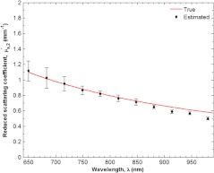
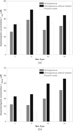
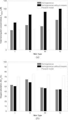
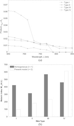
Monitoring of tissue blood volume and local oxygen saturation can inform the assessment of tissue health, healing, and dysfunction. These quantities can be estimated from the contribution of oxyhemoglobin and deoxyhemoglobin to the absorption spectrum of the dermis. However, estimation of blood related absorption in skin can be confounded by the strong absorption of melanin in the epidermis and epidermal thickness and pigmentation varies with anatomic location, race, gender, and degree of disease progression. Therefore, a method is desired that decouples the effect of melanin absorption in the epidermis from blood absorption in the dermis for a large range of skin types and thicknesses. A previously developed inverse method based on a neural network forward model was applied to simulated spatial frequency domain reflectance of skin for multiple wavelengths in the near infrared. It is demonstrated that the optical thickness of the epidermis and absorption and reduced scattering coefficients of the dermis can be determined independently and with minimal coupling. Then, the same inverse method was applied to reflectance measurements from a tissue simulating phantom and in vivo human skin. Oxygen saturation and total hemoglobin concentrations were estimated from the volar forearms of weakly and strongly pigmented subjects using a standard homogeneous model and the present two layer model.
Figures


Similar articles
-
Spatial frequency domain spectroscopy of two layer media.J Biomed Opt. 2011 Oct;16(10):107005. doi: 10.1117/1.3640814. J Biomed Opt. 2011. PMID: 22029367 Free PMC article.
-
Rapid and accurate estimation of blood saturation, melanin content, and epidermis thickness from spectral diffuse reflectance.Appl Opt. 2010 Apr 1;49(10):1707-19. doi: 10.1364/AO.49.001707. Appl Opt. 2010. PMID: 20357850
-
Determination of optical properties of turbid media spanning visible and near-infrared regimes via spatially modulated quantitative spectroscopy.J Biomed Opt. 2010 Jan-Feb;15(1):017012. doi: 10.1117/1.3299322. J Biomed Opt. 2010. PMID: 20210486 Free PMC article.
-
Hyperspectral imaging in diabetic foot wound care.J Diabetes Sci Technol. 2010 Sep 1;4(5):1099-113. doi: 10.1177/193229681000400508. J Diabetes Sci Technol. 2010. PMID: 20920429 Free PMC article. Review.
-
Skin optical properties in the obese and their relation to body mass index: a review.J Biomed Opt. 2022 Mar;27(3):030902. doi: 10.1117/1.JBO.27.3.030902. J Biomed Opt. 2022. PMID: 35352513 Free PMC article. Review.
Cited by
-
Single snapshot spatial frequency domain imaging for risk stratification of diabetes and diabetic foot.Biomed Opt Express. 2020 Jul 20;11(8):4471-4483. doi: 10.1364/BOE.394929. eCollection 2020 Aug 1. Biomed Opt Express. 2020. PMID: 32923057 Free PMC article.
-
Portable (handheld) clinical device for quantitative spectroscopy of skin, utilizing spatial frequency domain reflectance techniques.Rev Sci Instrum. 2017 Sep;88(9):094302. doi: 10.1063/1.5001075. Rev Sci Instrum. 2017. PMID: 28964218 Free PMC article.
-
Spatial frequency domain imaging in 2019: principles, applications, and perspectives.J Biomed Opt. 2019 Jun;24(7):1-18. doi: 10.1117/1.JBO.24.7.071613. J Biomed Opt. 2019. PMID: 31222987 Free PMC article. Review.
-
Modelling spatially-resolved diffuse reflectance spectra of a multi-layered skin model by artificial neural networks trained with Monte Carlo simulations.Biomed Opt Express. 2018 Mar 7;9(4):1531-1544. doi: 10.1364/BOE.9.001531. eCollection 2018 Apr 1. Biomed Opt Express. 2018. PMID: 29675300 Free PMC article.
-
Deep-learning-enabled spatial frequency domain imaging of the spatiotemporal dynamics of skin physiology.J Biomed Opt. 2025 Apr;30(4):046008. doi: 10.1117/1.JBO.30.4.046008. Epub 2025 Apr 20. J Biomed Opt. 2025. PMID: 40271202 Free PMC article.
References
-
- Bevilacqua F., et al. , “Method and apparatus for performing quantitative analysis and imaging surfaces and subsurfaces of turbid media using spatially structured illumination,” U.S. patent 6,958,815 B2 (2003).
