. 2012 Dec 4;109(49):19900-9.
doi: 10.1073/pnas.1116727109. Epub 2012 Oct 18.

Role of prefrontal cortex and the midbrain dopamine system in working memory updating

Affiliations

Role of prefrontal cortex and the midbrain dopamine system in working memory updating

Kimberlee D'Ardenne et al. Proc Natl Acad Sci U S A. .

Abstract

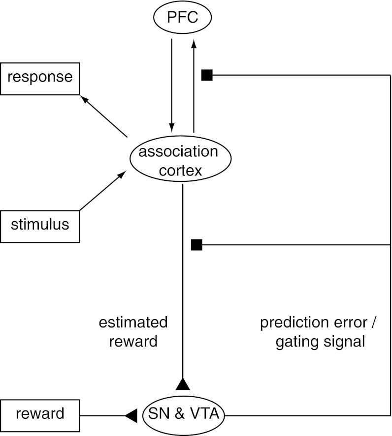
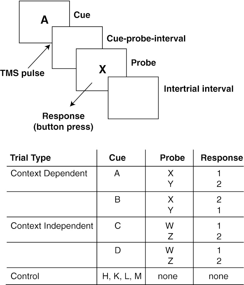
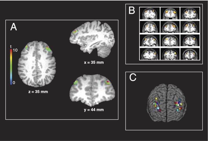
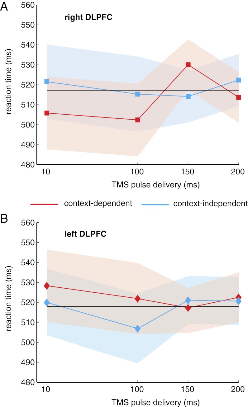
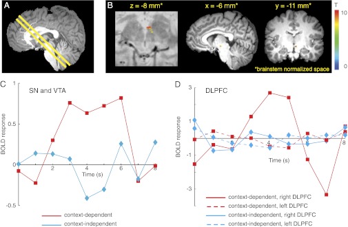
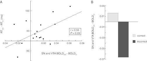
Humans are adept at switching between goal-directed behaviors quickly and effectively. The prefrontal cortex (PFC) is thought to play a critical role by encoding, updating, and maintaining internal representations of task context in working memory. It has also been hypothesized that the encoding of context representations in PFC is regulated by phasic dopamine gating signals. Here we use multimodal methods to test these hypotheses. First we used functional MRI (fMRI) to identify regions of PFC associated with the representation of context in a working memory task. Next we used single-pulse transcranial magnetic stimulation (TMS), guided spatially by our fMRI findings and temporally by previous event-related EEG recordings, to disrupt context encoding while participants performed the same working memory task. We found that TMS pulses to the right dorsolateral PFC (DLPFC) immediately after context presentation, and well in advance of the response, adversely impacted context-dependent relative to context-independent responses. This finding causally implicates right DLPFC function in context encoding. Finally, using the same paradigm, we conducted high-resolution fMRI measurements in brainstem dopaminergic nuclei (ventral tegmental area and substantia nigra) and found phasic responses after presentation of context stimuli relative to other stimuli, consistent with the timing of a gating signal that regulates the encoding of representations in PFC. Furthermore, these responses were positively correlated with behavior, as well as with responses in the same region of right DLPFC targeted in the TMS experiment, lending support to the hypothesis that dopamine phasic signals regulate encoding, and thereby the updating, of context representations in PFC.


Conflict of interest statement

The authors declare no conflict of interest.

Figures

Fig. 1.
Fig. 1.
Midbrain dopamine neurons in the SN and VTA broadcast signals that may be used to gate information into the PFC. Dopamine neurons have been shown to encode reward prediction errors through phasic changes in firing rate (25, 75). The reward prediction error is the difference between rewards received and rewards predicted (24). The theory that dopamine plays a role in updating working memory information in PFC (21) posits that the phasic increases in firing rate that encode reward prediction errors not only modulate the sensory inputs predicting reward (76, 77) but also simultaneously adjust the gain of inputs to the PFC (27). Modulation of these inputs permits the selective updating of representations in PFC. The reward prediction error and the gating signal work in concert because stimuli linked to the PFC representations needed to procure reward (e.g., representations of task rules and goals) themselves also predict reward. Arrows between PFC and association cortex indicate connectivity between cortical areas, triangles represent excitatory input to the SN and VTA, and squares represent gain modulation by dopamine at target sites.
Fig. 2.
Fig. 2.
Behavioral task. The AX-CPT task consisted of three types of trials: context-dependent, context-independent, and control. Control trials (not shown in the figure) consisted of display of a cue letter (500 ms) followed by an intertrial interval. Context-dependent and context-independent trials (shown) consisted of display of the cue (500 ms), a blank screen comprising the cue-probe interval, presentation of the probe (1,000 ms), the participant’s response to the probe, and finally the intertrial interval. Letters shown in the figure and table are examples (Materials and Methods gives details of actual stimulus assignments and interstimulus intervals). All responses involved pressing one of two buttons on a response box. In the TMS experiment, control trials were not included, and pulses were delivered at either 10, 100, 150, or 200 ms after the onset of the cue.
Fig. 3.
Fig. 3.
Whole-brain fMRI results and TMS stimulation sites for each participant. (A) Results of the group analysis of the fMRI data from experiment 1, identifying areas associated with context updating in the AX-CPT [n = 12; P < 0.05 corrected for multiple comparisons; left DLPFC region is 292 mm3 (9 contiguous voxels) and the right DLPFC region is 1199 mm3 (37 voxels)]. The statistical map is overlaid on the T1-weighted structural image in Talairach space from a representative participant. (B) Sites of activity in individual participants used to target TMS pulses in experiment 2. In all participants, this contrast identified activation in left and right DLPFC that was centered on BA 9 (n = 12; P < 10−6). Statistical maps for each participant are displayed on their T1-weighted structural images. (C) TMS stimulation sites in the right and left DLPFC for each participant in experiment 2. For each participant, the most active voxel within each area shown in B was chosen as the stimulation site. The locations of the stimulation sites are shown on a 3D reconstruction of a canonical Talairach brain. All images are displayed in radiological convention (left in the image corresponds to participant’s right side). Materials and Methods gives details of statistical analyses.
Fig. 4.
Fig. 4.
TMS disrupts performance during context-dependent trials. (A) TMS pulses applied to right DLPFC at 150 ms after cue onset significantly slowed reaction time compared with pulses applied at 10 ms after cue onset [t(11) = 2.26; P < 0.05] or at 100 ms [t(11) = 2.40; P < 0.05]. There was a nonsignificant trend at 200 ms [t(11) = 1.87; P < 0.1]. No other pulse time comparisons showed a significant effect on reaction time during context-dependent trials. (B) No pulse time comparisons showed a significant effect on reaction time during any trial type in left DLPFC. In A and B, shaded regions represent 95% confidence intervals, and the black line shows the overall mean reaction time.
Fig. 5.
Fig. 5.
BOLD responses in the midbrain SN and VTA were associated with context updating. (A) For brainstem fMRI, the midbrain was identified in the central sagittal slice of the T1-weighted structural image, and an oblique slab comprising axial/coronal slices was centered on as much of the SN and VTA as possible (constrained by the number of slices allowed by the participant's heart rate) and then tilted to include the regions of DLPFC shown in Fig. 3B. (B) A random effects general linear model analysis revealed that the BOLD response in the SN and VTA for context-dependent cues was greater than the BOLD response for context-independent cues (n = 24; P < 0.05, corrected for multiple comparisons). No other brain regions within the brainstem exhibited this effect. Statistical maps are overlaid on a proton-density weighted image (Left) and a T1-weighted image (Right) in brainstem-normalized space (74). (C) Average BOLD event-related time courses in SN and VTA (B) during context-dependent and context-independent trials in the AX-CPT task. The BOLD response to context-dependent cue letters (red) was greater than the BOLD responses to context-independent cue letters (blue). Cue letter presentation occurred at time t = 0. (D) Average BOLD event-related time courses from right and left DLPFC ROIs during context-dependent and context-independent trials. In right DLPFC, the BOLD response to context-dependent cue letters (solid red) was greater than the BOLD response to context-independent cue letters (solid blue). Left DLPFC (red and blue dashed lines) showed no BOLD responses to cue letters. BOLD time courses are in arbitrary MR units.
Fig. 6.
Fig. 6.
The SN and VTA BOLD responses to context updating were correlated with reaction time and were reduced on error trials. (A) Correlation across participants of differences in reaction time (RT) for context-dependent vs. context-independent trials with corresponding differences in the BOLD responses in SN and VTA (Fig. 5B). RT differences are in milliseconds; BOLD signal differences are in arbitrary MR units. (B) This same midbrain region exhibited significantly diminished context updating effect on BOLD response during error compared with correct trials [t(23) = 12.50; P = 10−12].

Comment in

  • Opening the gate to working memory.
    Badre D. Badre D. Proc Natl Acad Sci U S A. 2012 Dec 4;109(49):19878-9. doi: 10.1073/pnas.1216902109. Epub 2012 Nov 9. Proc Natl Acad Sci U S A. 2012. PMID: 23144220 Free PMC article. No abstract available.

References

    1. Duncan J. Disorganisation of behaviour after frontal lobe damage. Cogn Neuropsychol. 1986;3:271–290.
    1. Shallice T. From Neuropsychology to Mental Structure. Cambridge, UK: Cambridge Univ Press; 1988.
    1. Fuster JM, Alexander GE. Neuron activity related to short-term memory. Science. 1971;173(3997):652–654. - PubMed
    1. Goldman-Rakic PS, Leranth C, Williams SM, Mons N, Geffard M. Dopamine synaptic complex with pyramidal neurons in primate cerebral cortex. Proc Natl Acad Sci USA. 1989;86(22):9015–9019. - PMC - PubMed
    1. Wallis JD, Anderson KC, Miller EK. Single neurons in prefrontal cortex encode abstract rules. Nature. 2001;411(6840):953–956. - PubMed

Publication types