. 2012 Dec;13(12):1162-70.
doi: 10.1038/ni.2446. Epub 2012 Oct 21.

Control of RelB during dendritic cell activation integrates canonical and noncanonical NF-κB pathways

Affiliations

Control of RelB during dendritic cell activation integrates canonical and noncanonical NF-κB pathways

Vincent F-S Shih et al. Nat Immunol. 2012 Dec.

Abstract

The NF-κB protein RelB controls dendritic cell (DC) maturation and may be targeted therapeutically to manipulate T cell responses in disease. Here we report that RelB promoted DC activation not as the expected RelB-p52 effector of the noncanonical NF-κB pathway, but as a RelB-p50 dimer regulated by canonical IκBs, IκBα and IκBɛ. IκB control of RelB minimized spontaneous maturation but enabled rapid pathogen-responsive maturation. Computational modeling of the NF-κB signaling module identified control points of this unexpected cell type-specific regulation. Fibroblasts that we engineered accordingly showed DC-like RelB control. Canonical pathway control of RelB regulated pathogen-responsive gene expression programs. This work illustrates the potential utility of systems analyses in guiding the development of combination therapeutics for modulating DC-dependent T cell responses.


Figures

Figure 1
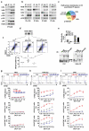
Figure 1. A MEF-based kinetic model does not account for RelB regulation in DCs
(a) Schematic of the distinct canonical and non-canonical NF-κB pathways identified in MEFs. Inflammatory signals lead to activation of the NEMO-containing kinase complex that triggers IκBα, -β, -ε degradation and the release of RelA-p50 into the nucleus. Developmental signals activate NIK–IKK1 kinase complex that results in p100 processing which allows for RelB-p52 nuclear translocation. (The IκBδ pathway is not shown for sake of clarity ). (b) Computational simulations using the MEF-based kinetic model version 5.0-MEF (see Supplementary Notes for details). The timecourse for nuclear RelA or RelB activity induced by LPS or LTβ stimulation are shown. (c) Quantitation of Rela, Relb and Nf-b2 transcript (left, by qRT-PCR) and of RelA, RelB, p100 and p52 protein (right, by immunoblot) numbers per cell in resting MEFs, BMDMs and BMDCs, graphed relative to the respective value in MEFs. (d) IKK1 and p52 abundance increase during DC differentiation. Whole cell extracts prepared from BMDC cell culture during a differentiation time course were subjected to IKK1 and p100/p52 immunoblotting. (e) in silico simulation of RelB cellular distribution using the mathematical model version 5.0-MEF describing NF-κB activation in MEFs as in Figure 1b or in model version 5.0-DC incorporating DC specific parameters derived from Figure 1c and 1d (see Supplementary Notes for details). (f) Bar graph showing quantitation of RelB molecules per WT BMDC distributed in cytoplasmic (CE) and nuclear (NE) fraction. Quantitation methods are described in Supplementary Fig. 1. (g) RelB, RelA and p100 immunoblots of cytoplasmic extracts prepared from the indicated cell types. Data shown are representative of at least three independent experiments (error bars, s.d.).
Figure 2
Figure 2. IκBα binding RelB-p50 limits spontaneous DC maturation
(a) RelB interacts with canonical IκBα and IκBε. RelB immunoprecipitates from whole DC extracts were probed for indicated interaction partners by immunoblotting. Fraction of proteins bound to RelB (IP) was compared to whole cell lysates (IN) and flow-through (FT). IgG immunoprecipitates served as an antibody specificity control. (b) IκBα and IκBε interact with RelB. IκB proteins were probed for their interaction with RelB using co-immunoprecipitation. Immunoblotting with other IκB family members served as negative controls. (c) NF-κB RelB DNA binding activities revealed by EMSA with nuclear extracts collected from Rel−/−Tnf−/−, Nfkbia−/−Tnf−/− Rel−/−, and Nfkbia−/−Tnf−/− Rel−/− Relb−/− BMDCs. (d) Quantitation of total RelB activity, RelB-p50 and RelB-p52 in immature BMDCs from indicated genotypes as revealed by EMSA. Band intensities of the antibody-ablation analysis (bottom) were summed and normalized to the total (panel c) and graphed relative to total RelB activity in WT BMDCs. (e) Constitutive expression of maturation markers is controlled by RelB. Fractions of CD11c+ BMDCs determined by FACS to be MHCIIhiCD86hi and MHCIIloCD86lo in indicated genotypes. Dot plots indicate the frequency of MHCIIhiCD86hi dendritic cells derived from GM-CSF-cultured Rel−/−Tnf−/− (n=5), Nfkbia−/−Tnf−/− Rel−/− (n=6), and Nfkbia−/−Tnf−/− Rel−/− Relb−/− (n=3) bone marrow cells in individual experiments. P*<0.01 (f-h) T-cell proliferation in DC:T cell co-cultures using Tnf−/−, Nfkbia−/−Tnf−/−, and Nfkbia−/− Tnf−/−Relb−/− BMDCs exposed to medium (f), OVA peptide (g) or OVA protein (h). Top, raw FACS data of CFSE-labeled T-cells stained for IL-2, showing proliferation-associated dye-dilution and IL-2 production. Middle, fraction of divided cells and, bottom, fraction of T-cells positive for the indicated activation-associated cytokine, graphed as a function of the DC:T cell ratio. Data shown in (a) (b) (c) (e) are representative of at least three independent experiments. Data in (f-h) are the average of duplicate leukocyte reactions produced for each of two independent BMDC cultures.
Figure 3
Figure 3. RelB-p50 is rapidly activated during TLR-mediated DC maturation
(a) Computational simulations of LPS-induced RelA and RelB activity during a 3 hour time course using the refined mathematical model version 5.1-DC. (b) NF-κB RelA (left) and NF-κB RelB (right) DNA binding activities monitored by EMSA. Nuclear extracts from WT BMDCs or WT MEFs activated by indicated stimuli were collected and subjected to EMSA. Equal amounts of nuclear proteins from BMDCs or MEFs were loaded and exposure of images was adjusted to reveal similar RelA peak activity in BMDCs and MEFs. (c) Computational simulations of RelB-p50 and RelB-p52 activities upon LPS stimulation that sum up to total nuclear RelB activity shown in panel A (top). Quantitation of RelB-p50 and RelB-p52 activities prior and after CpG stimulation were graphed relative to their respective basal activity (bottom). (d) IκB protein expression profiles induced by CpG. Whole cell extracts prepared from WT BMDCs were subjected to immunoblotting against antibodies as indicated. (e) Association of IκBα to RelB was monitored during a CpG time course by examining RelB immunoprecipitates from CpG-stimulated WT BMDCs. Immunoprecipitation with Relb−/− extracts (C) serves as a control, indicating specificity of RelB antibody. (f) Computational simulations of CpG-induced RelB activation in mathematical models, based on version 5.1-DC that were deficient in the indicated proteins. (g) CpG-induced NF-κB RelB DNA binding activities in indicated gene-deficient BMDCs were monitored by EMSA (top). Signals were quantitated and graphed relative to respective resting cells (bottom). Data shown in (b) (d) (e) (g) are representative of at least three independent experiments. Data shown in (c) is representative of two independent experiments (error bars: s.d.)
Figure 4
Figure 4. Determinants of RelB’s responsiveness to canonical signals
(a) Heatmap depicting how LPS-inducibility of RelB is a function RelB synthesis and NIK halflife. The results derived from in silico simulations of peak nuclear RelB-p50 DNA abundance (nM) during an LPS time course when modulating the halflife of NIK (y-axis) and the mRNA synthesis rate of RelB (x-axis). (b) Immunoblots with indicated antibodies of whole cell extracts collected from control or Traf3−/− MEFs reconstituted with empty vector (EV) or RelB transgene (RelB-TG). The top band in the RelB blot represents exogenous protein whereas the bottom band represents endogenous RelB protein. (c) NF-κB RelB (top) and RelA (bottom) DNA binding activities induced by LPS were monitored with nuclear extracts collected from control or Traf3−/− MEFs transduced with empty vector (EV) or a RelB transgene (RelB-TG). (d) Quantitation of RelB-p50 and RelB-p52 activities in LPS-stimulated Traf3−/−(RelB-Tg) MEFs; signals were graphed relative to respective RelB-containing dimers basal activity. (e) Single cell data at indicated time points of the nuclear localization of a retrovirally expressed RelB-GFP fusion protein in response to TNF stimulation of control or Traf3−/− MEFs. (f) Schematic depicting the regulation of RelB by non-canonical or canonical stimuli. RelB may either dimerize with p52 in response to stimulus-induced non-canonical stimuli, or dimerize with p50 and become responsive to canonical stimuli. Cell-type-specific steady-state control of RelB expression and non-canonical pathway activity determines which stimuli activate RelB: at low steady-state levels, RelB is responsive to non-canonical stimuli as reported in MEFs; at high steady-state levels RelB will dimerize not only p52 but also p50, and becomes responsive to canonical stimuli via IκBα and IκBε control. Data shown here are representative of two independent experiments (error bars: s.d.).
Figure 5
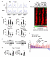
Figure 5. RelB regulates DC activation markers and inflammatory mediators
(a) Analysis of cell surface maker expression in WT and Relb−/− BMDCs in response to CpG. Cells untreated (grey) or treated with CpG (blue) or Pam3CSK4 (red) for 24 hours were subjected to FACS analysis. (b) Gene expression analyses of WT and Relb−/− BMDCs stimulated with CpG or Pam3CSK4 for the indicated time course by qRT-PCR. Signals were graphed relative to respective resting cells. (c) EMSA with nuclear extracts harvested from CpG-stimulated WT BMDCs using DNA probes containing the κB-site containing promoter sequence from Tnf or Il23a gene. (d) Chromatin immunoprecipitation analyses with RelB or IgG control antibodies using cell extracts from WT BMDCs collected prior or 75 minutes after stimulation with CpG. Quantitation of DNA precipitated was performed by qPCR with primers corresponding to the promoter region of indicated genes and graphed relative to input signals. (e) Microarray mRNA expression analysis from WT and Rel−/−Relb−/− BMDCs stimulated with CpG and Pam3CSK4 for indicated time points. Heatmap showing the expression pattern from one experiment in a (log2) fold induction scale of 157 significant down-regulated genes in Rel−/−Relb−/− BMDCs identified by Significant Analysis of Microarray (SAM). Color scale “1.0” denotes normalized highest expression value of the given gene across time courses. (f) RelB and c-Rel regulate overlapping sets of genes. The expression phenotype caused by RelB-deficiency was determined for the 50 genes with the most severe expression defect in Rel−/−Relb−/− BMDCs. The list of genes was sorted expression difference between WT and Relb−/− BMDCs. Data shown in (a) (b) (c) (d) are representative of at least three independent experiments (error bars: s.e.m.). *P<0.05, **P<0.01.
Figure 6
Figure 6. RelB may mediate cRel functions in DCs
(a) Immunoblot for RelA, RelB and cRel of whole cell extracts prepared from indicated gene-deficient BMDCs. α-tubulin serves as loading control. (b) Amount of Relb transcripts was compared by qRT-PCR with mRNA collected from wild type and Rel−/− BMDCs and graphed relative to WT cells. (c) NF-κB DNA binding activities of RelB, c-Rel and RelA induced by LPS in indicated gene-deficient BMDCs were monitored by EMSA. Data shown are representative of three independent experiments (error bars: s.e.m.). *P<0.01.

Comment in

References

    1. Banchereau J, Steinman RM. Dendritic cells and the control of immunity. Nature. 1998;392:245–252. - PubMed
    1. Liu YJ. IPC: professional type 1 interferon-producing cells and plasmacytoid dendritic cell precursors. Annu Rev Immunol. 2005;23:275–306. - PubMed
    1. Serbina NV, Salazar-Mather TP, Biron CA, Kuziel WA, Pamer EG. TNF/iNOS-producing dendritic cells mediate innate immune defense against bacterial infection. Immunity. 2003;19:59–70. - PubMed
    1. Steinman RM, Banchereau J. Taking dendritic cells into medicine. Nature. 2007;449:419–426. - PubMed
    1. Carrasco D, Ryseck RP, Bravo R. Expression of relB transcripts during lymphoid organ development: specific expression in dendritic antigen-presenting cells. Development. 1993;118:1221–1231. - PubMed

Publication types

MeSH terms

Associated data