. 2012 Dec;13(12):1213-21.
doi: 10.1038/ni.2454. Epub 2012 Oct 21.

The CD46-Jagged1 interaction is critical for human TH1 immunity

Affiliations

The CD46-Jagged1 interaction is critical for human TH1 immunity

Gaëlle Le Friec et al. Nat Immunol. 2012 Dec.

Abstract

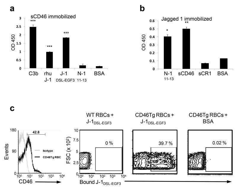
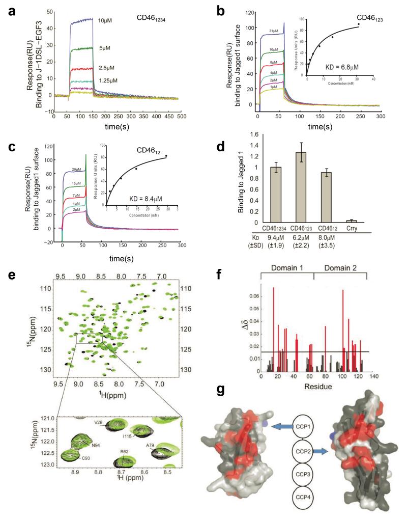
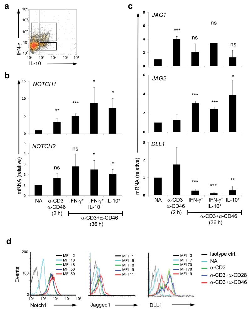
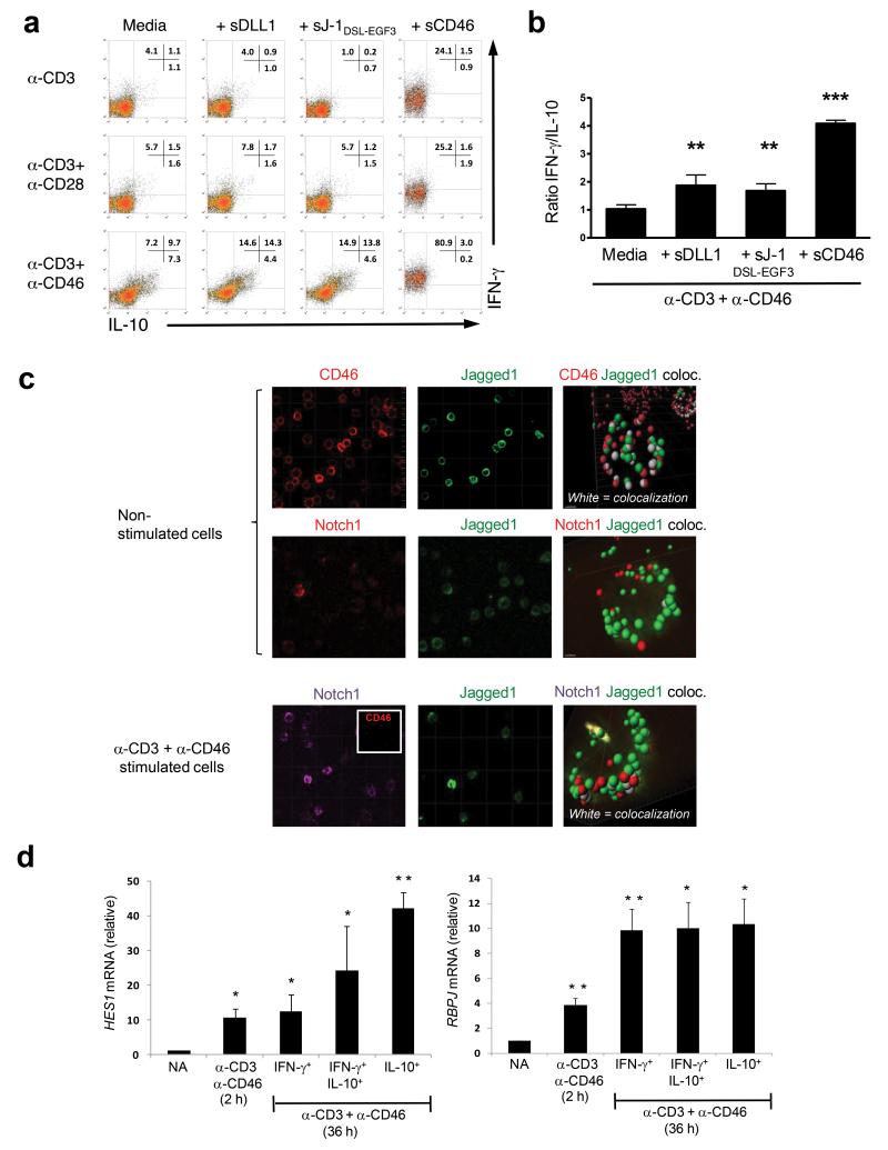
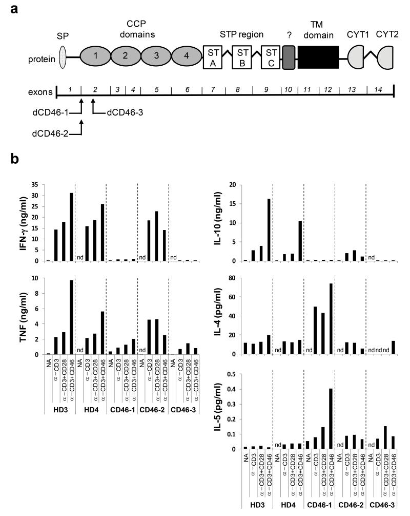
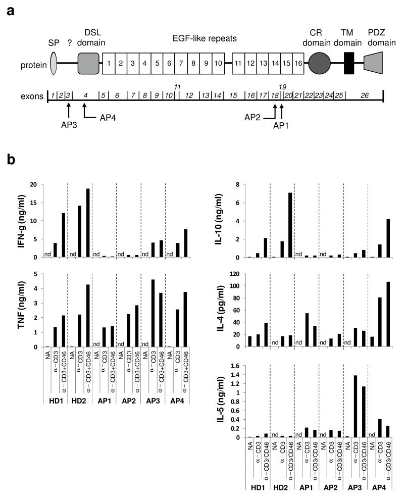
CD46 is a complement regulator with important roles related to the immune response. CD46 functions as a pathogen receptor and is a potent costimulator for the induction of interferon-γ (IFN-γ)-secreting effector T helper type 1 (T(H)1) cells and their subsequent switch into interleukin 10 (IL-10)-producing regulatory T cells. Here we identified the Notch family member Jagged1 as a physiological ligand for CD46. Furthermore, we found that CD46 regulated the expression of Notch receptors and ligands during T cell activation and that disturbance of the CD46-Notch crosstalk impeded induction of IFN-γ and switching to IL-10. Notably, CD4(+) T cells from CD46-deficient patients and patients with hypomorphic mutations in the gene encoding Jagged1 (Alagille syndrome) failed to mount appropriate T(H)1 responses in vitro and in vivo, which suggested that CD46-Jagged1 crosstalk is responsible for the recurrent infections in subpopulations of these patients.


Figures

Figure. 1
Figure. 1. Jagged1 is a ligand for CD46
a and b, Analysis of the CD46 and Jagged1 interaction by ELISA. ELISA assays were performed utilizing either (a) immobilized soluble CD46 (CCP1-4) incubated with indicated recombinant soluble proteins (rhu J-1, recombinant human Jagged1; J-1DSL-EGF3, a recombinant protein containing the Notch1 binding site DSL and the first three EGF-like domains; N-111-13, a recombinant protein containing Notch1 EGF-like domains 11-13) or bovine serum albumin (BSA) or (b) immobilized J-1DSL-EGF3 incubated with indicated soluble proteins (sCR1, soluble recombinant complement receptor 1) in Ca2+ buffer (for further details on utilized proteins, see Supplementary Fig. 1). Mean ± SD (n = 5). (c) Analysis of Jagged1 binding to cell surface-expressed CD46. Red blood cells (RBCs) from CD46Tg mice express CD46 on the surface (left panel). RBCs from CD46Tg and from WT mice were incubated with biotinylated J-1DSL-EGF3 (middle panels) or BSA (right panel) and J-1DSL-EGF3-binding to RBCs measured using fluorochrome-labeled streptavidin. Shown is one representative data set of three independently performed experiments. *, p < 0.05; **, p < 0.005; ***, p < 0.001 when compared with BSA binding to immobilized protein.
Figure. 2
Figure. 2. Jagged1 binds to CD46 CCPs 1 and 2
(a) Surface plasmon resonance demonstrates that J-1DSL-EGF3, binds to CD461-4 coupled on the surface of the chip with a KD of ~8μM (normalised by subtraction of mock-coupled channel). (b and c) Binding of (b) CD461-3 and (c) CD461-2 to immobilized J-1DSL-EGF3 normalized by subtraction of a mock-coupled channel. Insert chart shows equilibrium values of binding and KD fit using SigmaPlot. All data processed using the BIAevaluation software. (d) Injections of CD461-4 (21 μM), CD461-3 (25 μM) CD461-2 (19 μM) over immobilized J-1DSL-EGF3 in triplicate. The rodent complement regulator Crry (CCP1-4) was used as a negative control (20μM). Data are normalised for construct molecular weight and represented as mean ± SD. The KD values reported below the chart are derived from a minimum of three repeated dilution series ± SD. (e) NMR spectroscopy overlay of the 1H,15N-heteronuclear single quantum coherence (HSQC) of CD461-2 (black) demonstrates chemical shift perturbation with the addition of unlabelled J-1DSL-EGF3 (green) at a molar ratio of 0.6. (f) Chemical shift perturbation by residue for those unambiguously assigned and baseline resolved in the 15N,1H-HSQC for CD461-2. (g) Surface structure of CCP1 and CCP2 (3O8E.pdb) showing residues with unambiguous assignment (dark grey) and chemical shift perturbation > 0.15ppm (red) upon J-1DSL-EGF3 addition. N-linked glycosylation sites are shown in blue.
Figure. 3
Figure. 3. CD46 regulates Notch receptors and ligand expression on human CD4+ T cells
(a) CD3+CD46+IL-2 activation of human CD4+ T cells induces three distinct IFN-γ and IL-10-secreting subpopulations (36h post activation). (b and c) CD46 co-stimulation increases mRNA levels of NOTCH1, NOTCH2 and JAG1 and JAG2 genes but decreases DLL1 transcripts. mRNAs were assessed from non-activated T cells (NA), 2h α-CD3+CD46-activated T cells (with 50 U/ml rhIL-2) and from T cells corresponding to the three distinct cytokine-secreting populations. Expression of mRNA is presented relative to 18s mRNA expression in each sample. Results shown are mean ± SD (n = 4). mRNAs coding for NOTCH3 and NOTCH4 as well as DLL4 genes were present but unaltered by CD3 or CD3+CD46 activation at any time point measured (data not shown). (d) Protein expression of Notch1, Jagged1 and Delta-like1 (DLL1) on non-subsorted bulk CD4+ T cells after CD3+CD46 stimulation for 36h. Data represent mean ± SD (n=4). Note that increase in proteins also reached statistical significance when the respective mean fluorescence intensities (MFIs) of the histograms were compared. *, p < 0.05; **, p < 0 .005; ***, p < 0.001 when compared with non-activated cells. Ctrl, control; ns, statistically not significant.
Figure. 4
Figure. 4. Undisturbed CD46 and Notch system crosstalk is required for normal IFN-γ to IL-10 switching in human TH1 cells
(a) Addition of sDLL1, sJ-1DSL-EGF3 or sCD46 during CD3, CD3+CD28 or CD3+CD46 activation of CD4+ T cells increases the numbers of IFN-γ-secreting TH1 cells and (b) changes the IFN-γ:IL-10 ratio of cytokines secreted into the media. Results shown are mean ± SD (n = 6). (c) CD46 sequesters Jagged1 on resting T cells and prevents the Jagged1 and Notch1 interaction. Non-activated (upper two rows of panels) or CD3+CD46-activated (lower row of panels) T cells were stained with antibodies to CD46, Notch1 or Jagged1 and then subjected to superresolution confocal microscopy and 3D analysis to assess for molecular co-localization. White areas in the 3D analyses indicate co-localization of assessed proteins. CD3+CD46-activated T cells lose CD46 surface expression and become negative for CD46 staining (see insert in left panel, lower row). Shown are representative results of two independently performed experiments. (d) CD46 co-stimulation of purified CD4+ T cells increases HES1 and RBPJ mRNA transcription. T cells were activated as indicated and the three emerging IFN-γ- and IL-10-secreting populations from the 36hr sample cell sorted. mRNA was purified and then subjected to quantitative PCR analysis for HES1 and RBPJ transcripts. Expression of mRNA is presented relative to 18s mRNA expression in each sample. Data represent mean ± SD (n=3). *, p < 0.05 **, p < 0.005, ***, p < 0 .001 when compared to media (b) or non-activated cells (d).
Figure. 5
Figure. 5. T cells from CD46-deficient patients present with defective in vitro TH1 induction
(a) Localization of the CD46 gene mutations in the three CD46-deficient patients (CD46-1 to CD46-3) assessed for TH1 induction. The lower part shows the exon structure of the CD46 gene and above the corresponding protein domains. (b) Comparison of cytokine expression by CD4+ T cells from healthy donors and CD46-deficient patients. T cells from two healthy donors (HD3 and HD4) or patients were purified from freshly-drawn blood samples and either left non-activated (NA), or activated with immobilized antibodies to CD3 and CD46 in the presence of 25 U/ml rhIL-2. Indicated cytokine secretion into the cell culture media was assessed 36h post activation using the TH1 and TH2 CBA Cytokine Secretion Assay. Data shown are the mean value of each condition performed in duplicate. Note, that though values for only two HD are shown, these are representative for 12 age- and gender-matched donors assessed over the course of the study. CCP, complement control protein; CYT1 or CYT2, cytoplasmic tail 1 or −2; nd, not detectable; SP, signal peptide; STP, serine threonine proline-rich; TM, transmembrane;?, region of unknown function
Figure. 6
Figure. 6. T cells from Alagille Syndrome patients present with defective in vitro TH1 induction
(a) Localization of the JAG1 gene mutations in the four Alagille Syndrome patients (AP1 to AP4) assessed for TH1 induction. The lower part depicts the exon structure of the JAG1 gene with exons 1 to 26 and the upper part the corresponding Jagged1 protein domains. Note that all four JAG1 mutations result likely in the retention of the altered protein in the endoplasmic reticulum. (b) Comparison of cytokine expression by CD4+ T cells from healthy donors and APs. T cells from two healthy donors (HD1 and HD2) or APs1-4 were purified from freshly-drawn blood samples and either left non-activated (NA), or activated with immobilized antibodies to CD3 and CD46 in the presence of 25 U/ml rhIL-2. Indicated cytokine secretion into the cell culture media was assessed 36h post activation using the TH1 and TH2 CBA Cytokine Secretion Assay. Data shown are the mean value of each condition performed in duplicate. CR, cysteine rich; nd, not detectable; PDZ, post synaptic density protein (PSD95), Drosophila disc large tumor suppressor (Dlg1), and zonula occludens-1 protein (zo-1); SP, signal peptide; TM, transmembrane;?, region of unknown function

Comment in

References

    1. Liszewski MK, Post TW, Atkinson JP. Membrane cofactor protein (MCP or CD46): newest member of the regulators of complement activation gene cluster. Annu Rev Immunol. 1991;9:431–455. - PubMed
    1. Cattaneo R. Four viruses, two bacteria, and one receptor: membrane cofactor protein (CD46) as pathogens’ magnet. J Virol. 2004;78:4385–4388. - PMC - PubMed
    1. Astier A, Trescol-Biemont MC, Azocar O, Lamouille B, Rabourdin-Combe C. Cutting edge: CD46, a new costimulatory molecule for T cells, that induces p120CBL and LAT phosphorylation. J Immunol. 2000;164:6091–6095. - PubMed
    1. Cardone J, et al. Complement regulator CD46 temporally regulates cytokine production by conventional and unconventional T cells. Nat Immunol. 2010;11:862–871. - PMC - PubMed
    1. Cope A, Le Friec G, Cardone J, Kemper C. The Th1 life cycle: molecular control of IFN-gamma to IL-10 switching. Trends Immunol. 2011;32:278–286. - PubMed

Publication types

MeSH terms