

Polyomavirus JC infection inhibits differentiation of oligodendrocyte progenitor cells
- PMID: 23086711
- PMCID: PMC4641310
- DOI: 10.1002/jnr.23135
Polyomavirus JC infection inhibits differentiation of oligodendrocyte progenitor cells
Abstract
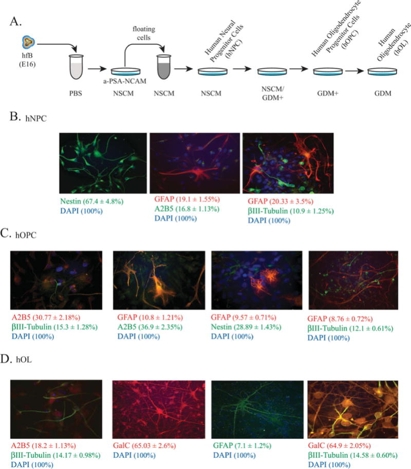
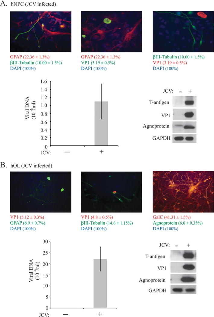
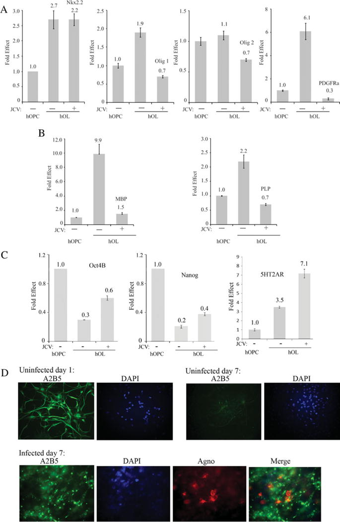
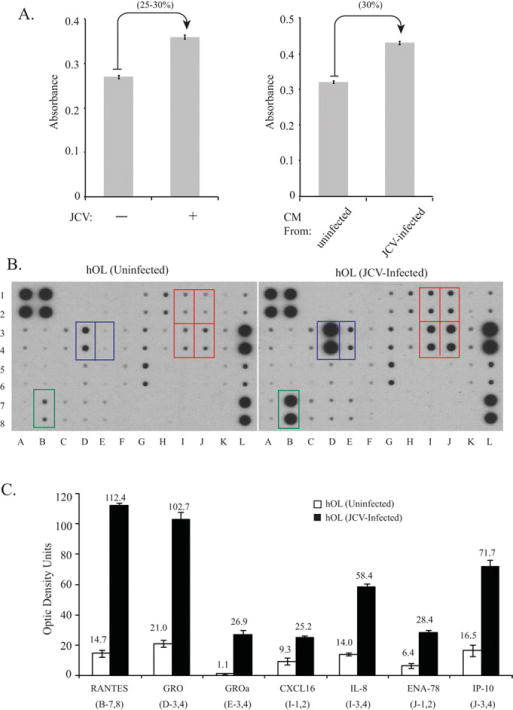
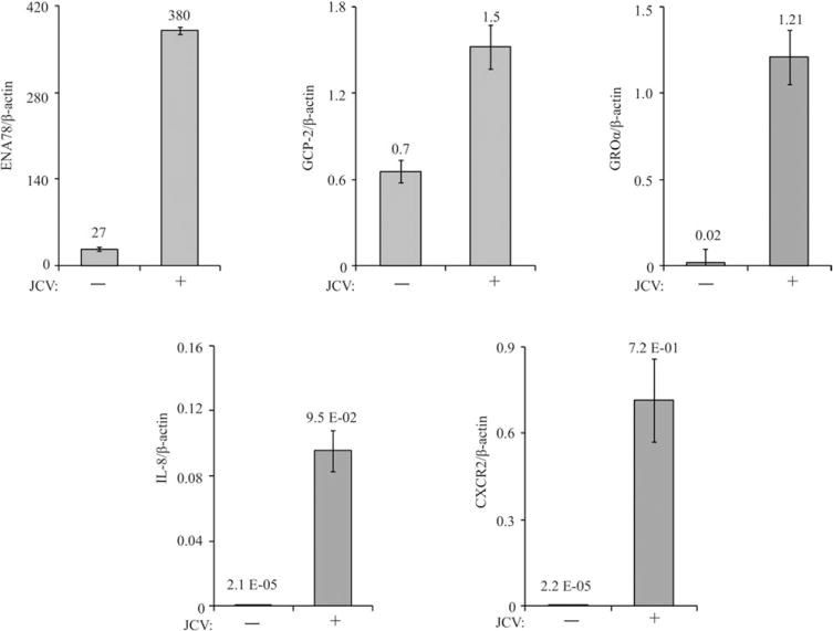
Reactivation of the human polyomavirus JC (JCV) in the CNS results in a fatal demyelinating disease, progressive multifocal leukoencephalopathy (PML). The lytic destruction of oligodendrocytes, which occurs at the terminal stage of the viral infection cycle, is considered a critical factor in the development of demyelination and the pathogenesis of PML. However, knowledge is limited about interaction of JCV with oligodendrocytes and its impact on the denudation of axons at the early stage of viral reactivation and prior to the destruction of the infected cells. We have developed an in vitro neuroprogenitor cell culture using human fetal brain that can be differentiated to the oligodendrocyte lineage to investigate interactions of JCV with its host cells. Results show that infection with JCV delays oligodendrocyte maturation as shown by reduced levels of oligodendrocytic markers, including myelin basic protein, proteolipid protein, and platelet-derived growth factor receptor-α. Furthermore, replication of JCV in these cells caused substantial dysregulation of several chemokines, including CCL5/RANTES, GRO, CXCL1/GROα, CXCL16, CXCL8/IL-8, CXCL5/ENA-78, and CXCL10/IP-10, all of which play a role in cell growth and differentiation.
Copyright © 2012 Wiley Periodicals, Inc.
Figures
Similar articles
-
New JC virus infection patterns by in situ polymerase chain reaction in brains of acquired immunodeficiency syndrome patients with progressive multifocal leukoencephalopathy.J Neurovirol. 2004 Feb;10(1):1-11. doi: 10.1080/13550280490269691. J Neurovirol. 2004. PMID: 14982723
-
Replication of JC Virus DNA in the G144 Oligodendrocyte Cell Line Is Dependent Upon Akt.J Virol. 2017 Sep 27;91(20):e00735-17. doi: 10.1128/JVI.00735-17. Print 2017 Oct 15. J Virol. 2017. PMID: 28768870 Free PMC article.
-
Effects of JC virus infection on anti-apoptotic protein survivin in progressive multifocal leukoencephalopathy.Am J Pathol. 2007 Apr;170(4):1291-304. doi: 10.2353/ajpath.2007.060689. Am J Pathol. 2007. PMID: 17392168 Free PMC article.
-
Traffic of JC virus from sites of initial infection to the brain: the path to progressive multifocal leukoencephalopathy.J Infect Dis. 2002 Dec 1;186 Suppl 2:S180-6. doi: 10.1086/344280. J Infect Dis. 2002. PMID: 12424695 Review.
-
A review on JC virus infection in kidney transplant recipients.Clin Dev Immunol. 2013;2013:926391. doi: 10.1155/2013/926391. Epub 2013 Jan 29. Clin Dev Immunol. 2013. PMID: 23424601 Free PMC article. Review.
Cited by
-
Molecular Markers in Maternal Blood Exosomes Allow Early Detection of Fetal Alcohol Spectrum Disorders.Int J Mol Sci. 2022 Dec 21;24(1):135. doi: 10.3390/ijms24010135. Int J Mol Sci. 2022. PMID: 36613580 Free PMC article.
-
Inflammation and Elevated Osteopontin in Plasma and CSF in Cerebral Malaria Compared to Plasmodium-Negative Neurological Infections.Int J Mol Sci. 2024 Sep 5;25(17):9620. doi: 10.3390/ijms25179620. Int J Mol Sci. 2024. PMID: 39273566 Free PMC article.
-
Effect of the Large and Small T-Antigens of Human Polyomaviruses on Signaling Pathways.Int J Mol Sci. 2019 Aug 12;20(16):3914. doi: 10.3390/ijms20163914. Int J Mol Sci. 2019. PMID: 31408949 Free PMC article. Review.
-
CC and CXC chemokines play key roles in the development of polyomaviruses related pathological conditions.Virol J. 2021 Jun 3;18(1):111. doi: 10.1186/s12985-021-01582-4. Virol J. 2021. PMID: 34082771 Free PMC article. Review.
-
Progressive multifocal leukoencephalopathy following five lines of therapy and three autologous bone marrow transplants for multiple myeloma.BMJ Case Rep. 2020 Mar 22;13(3):e233552. doi: 10.1136/bcr-2019-233552. BMJ Case Rep. 2020. PMID: 32205382 Free PMC article.
References
-
- Alvarez-Buylla A, Kohwi M, Nguyen TM, Merkle FT. The heterogeneity of adult neural stem cells and the emerging complexity of their niche. Cold Spring Harbor Symp Quant Biol. 2008;73:357–365. - PubMed
-
- Bajetto A, Bonavia R, Barbero S, Schettini G. Characterization of chemokines and their receptors in the central nervous system: physiopathological implications. J Neurochem. 2002;82:1311–1329. - PubMed
-
- Bartosik-Psujek H, Stelmasiak Z. The levels of chemokines CXCL8, CCL2 and CCL5 in multiple sclerosis patients are linked to the activity of the disease. Eur J Neurol. 2005;12:49–54. - PubMed
-
- Berger JR. Progressive multifocal leukoencephalopathy and newer biological agents. Drug Saf. 2010;33:969–983. - PubMed
Publication types
MeSH terms
Substances
Grants and funding
LinkOut - more resources
Full Text Sources
