

Correlation of urine TMPRSS2:ERG and PCA3 to ERG+ and total prostate cancer burden
- PMID: 23086769
- PMCID: PMC3597433
- DOI: 10.1309/AJCPU7PPWUPYG8OH
Correlation of urine TMPRSS2:ERG and PCA3 to ERG+ and total prostate cancer burden
Abstract
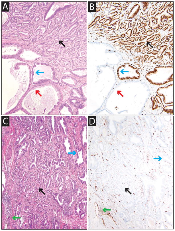
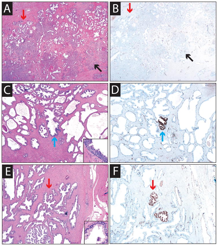
ERG rearrangements (most commonly transmembrane protease, serine 2 [TMPRSS2]:ERG [T2:ERG] gene fusions) have been identified in approximately 50% of prostate cancers . Quantification of T2:ERG in postdigital rectal examination urine, in combination with PCA3, improves the performance of serum prostate-specific antigen for prostate cancer prediction on biopsy. Here we compared urine T2:ERG and PCA3 scores with ERG+ (determined with immunohistochemical analysis) and total prostate cancer burden in 41 mapped prostatectomies. Prostatectomies had a median of 3 tumor foci (range, 1-15) and 2.6 cm of summed linear tumor dimension (range, 0.6-7.1 cm). Urine T2:ERG score correlated most with summed linear ERG+ tumor dimension and number of ERG+ foci (r(s) = 0.68 and 0.67, respectively, both P < .001). Urine PCA3 score showed weaker correlation with both number of tumor foci (r(s) = 0.34, P = .03) and summed linear tumor dimension (r(s) = 0.26, P = .10). In summary, we demonstrate a strong correlation between urine T2:ERG score and total ERG+ prostate cancer burden at prostatectomy, consistent with high tumor specificity.
Conflict of interest statement
The University of Michigan has been issued a patent on the detection of ETS gene fusions in prostate cancer, on which A.M.C. and S.A.T. are listed as co-inventors. The University of Michigan licensed the diagnostic field of use to Gen-Probe, Inc, which sublicensed rights to Ventana Medical Systems, Inc. Neither company played a role in data collection, interpretation or analysis, and did not participate in the study design or the decision to submit for publication. N.P. has served as a consultant for Ventana Medical Systems. A.M.C. has served as consultant to Gen-Probe, Inc. and Ventana Medical Systems. S.A.T. has received honoraria from Ventana Medical Systems.
Figures
Similar articles
-
Urine TMPRSS2:ERG Plus PCA3 for Individualized Prostate Cancer Risk Assessment.Eur Urol. 2016 Jul;70(1):45-53. doi: 10.1016/j.eururo.2015.04.039. Epub 2015 May 16. Eur Urol. 2016. PMID: 25985884 Free PMC article.
-
Urine TMPRSS2:ERG fusion transcript integrated with PCA3 score, genotyping, and biological features are correlated to the results of prostatic biopsies in men at risk of prostate cancer.Prostate. 2013 Feb 15;73(3):242-9. doi: 10.1002/pros.22563. Epub 2012 Jul 20. Prostate. 2013. PMID: 22821767
-
Prospective multicentre evaluation of PCA3 and TMPRSS2-ERG gene fusions as diagnostic and prognostic urinary biomarkers for prostate cancer.Eur Urol. 2014 Mar;65(3):534-42. doi: 10.1016/j.eururo.2012.11.014. Epub 2012 Nov 15. Eur Urol. 2014. PMID: 23201468
-
Urinary Biomarkers for Prostate Cancer.Urol Clin North Am. 2016 Feb;43(1):17-38. doi: 10.1016/j.ucl.2015.08.003. Urol Clin North Am. 2016. PMID: 26614026 Review.
-
Prostate Cancer Detection and Prognosis: From Prostate Specific Antigen (PSA) to Exosomal Biomarkers.Int J Mol Sci. 2016 Oct 26;17(11):1784. doi: 10.3390/ijms17111784. Int J Mol Sci. 2016. PMID: 27792187 Free PMC article. Review.
Cited by
-
Preliminary Results of Noninvasive Detection of TMPRSS2:ERG Gene Fusion in a Cohort of Patients With Localized Prostate Cancer.Korean J Urol. 2013 Jun;54(6):359-63. doi: 10.4111/kju.2013.54.6.359. Epub 2013 Jun 12. Korean J Urol. 2013. PMID: 23789042 Free PMC article.
-
Gene-Transcript Expression in Urine Supernatant and Urine Cell-Sediment Are Different but Equally Useful for Detecting Prostate Cancer.Cancers (Basel). 2023 Jan 27;15(3):789. doi: 10.3390/cancers15030789. Cancers (Basel). 2023. PMID: 36765747 Free PMC article.
-
Assessment of long-term outcomes associated with urinary prostate cancer antigen 3 and TMPRSS2:ERG gene fusion at repeat biopsy.Cancer. 2015 Nov 15;121(22):4071-9. doi: 10.1002/cncr.29611. Epub 2015 Aug 17. Cancer. 2015. PMID: 26280815 Free PMC article.
-
Significance of the TMPRSS2:ERG gene fusion in prostate cancer.Mol Med Rep. 2017 Oct;16(4):5450-5458. doi: 10.3892/mmr.2017.7281. Epub 2017 Aug 18. Mol Med Rep. 2017. PMID: 28849022 Free PMC article.
-
Molecular Profiling to Determine Clonality of Serial Magnetic Resonance Imaging/Ultrasound Fusion Biopsies from Men on Active Surveillance for Low-Risk Prostate Cancer.Clin Cancer Res. 2017 Feb 15;23(4):985-991. doi: 10.1158/1078-0432.CCR-16-1454. Epub 2016 Oct 7. Clin Cancer Res. 2017. PMID: 28031426 Free PMC article.
References
-
- Tomlins SA, Rhodes DR, Perner S, et al. Recurrent fusion of TMPRSS2 and ETS transcription factor genes in prostate cancer. Science. 2005;310:644–648. - PubMed
-
- Rosen P, Sesterhenn IA, Brassell SA, et al. Clinical potential of the ERG oncoprotein in prostate cancer. Nat Rev Urol. 2012 - PubMed
-
- Tomlins SA, Mehra R, Rhodes DR, et al. Integrative molecular concept modeling of prostate cancer progression. Nat Genet. 2007;39:41–51. - PubMed
Publication types
MeSH terms
Substances
Grants and funding
LinkOut - more resources
Full Text Sources
Medical
