Comparative Study
. 2012 Dec;18(12):2128-34.
doi: 10.1261/rna.034967.112. Epub 2012 Oct 19.

Crystal structure of the primary piRNA biogenesis factor Zucchini reveals similarity to the bacterial PLD endonuclease Nuc

Comparative Study

Crystal structure of the primary piRNA biogenesis factor Zucchini reveals similarity to the bacterial PLD endonuclease Nuc

Franka Voigt et al. RNA. 2012 Dec.

Abstract

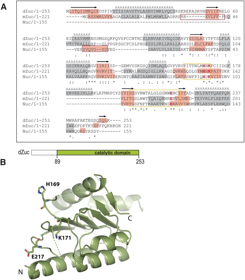
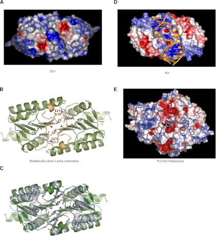
Piwi-interacting RNAs (piRNAs) are a gonad-specific class of small RNAs that associate with the Piwi clade of Argonaute proteins and play a key role in transposon silencing in animals. Since biogenesis of piRNAs is independent of the double-stranded RNA-processing enzyme Dicer, an alternative nuclease that can process single-stranded RNA transcripts has been long sought. A Phospholipase D-like protein, Zucchini, that is essential for piRNA processing has been proposed to be a nuclease acting in piRNA biogenesis. Here we describe the crystal structure of Zucchini from Drosophila melanogaster and show that it is very similar to the bacterial endonuclease, Nuc. The structure also reveals that homodimerization induces major conformational changes assembling the active site. The active site is situated on the dimer interface at the bottom of a narrow groove that can likely accommodate single-stranded nucleic acid substrates. Furthermore, biophysical analysis identifies protein segments essential for dimerization and provides insights into regulation of Zucchini's activity.


Figures

FIGURE 1.
FIGURE 1.
Structure of Drosophila Zucchini. (A) Sequence alignment of Drosophila Zuc (dZuc), mouse Zuc (mZuc), and the bacterial Nuc. Sequence conservation is shown below the alignment. Active site loops that are disordered in the dZuc monomer structure are marked with a yellow box, the catalytic residues shown in the structure figures are highlighted (H, K, E in red), α-helices are shown with gray background, while β-strands are highlighted in orange. The N-terminal β-strand of Nuc (red box) is missing from the Zuc proteins; its role in dimer formation is probably exerted by residues 36–44 in mZuc (note predicted β-strand, red box). (B) The cartoon indicates the dZuc construct used for crystallization. Crystal structure of dZuc is shown in ribbon representation and catalytic residues are shown as sticks. Green dashed lines connect loops with missing density.
FIGURE 2.
FIGURE 2.
Zucchini forms homodimers. (A) Crystal structure of dimeric Nuc (PDB 1BYR) (Stuckey and Dixon 1999). The N-terminal β-strand (dark red) is required for dimerization. The active site loops (yellow) are in their catalytically competent conformation. (B) dZuc dimer modeled on the related bacterial nuclease Nuc dimer structure. The active site loops (yellow) are misplaced and disordered resulting in clashes on the dimer interface (red circles). (C) Gel-filtration chromatograms for three mouse Zucchini (mZuc) constructs. Dimer formation, as determined by elution volumes (in milliliters), is seen only with the construct mZuc(36–221 aa). (D) Analytical ultracentrifugation analysis of mZuc shows that it sediments as a dimer.
FIGURE 3.
FIGURE 3.
Zucchini dimer probably accepts ssRNA substrates. (A) Surface representation of dZuc dimer colored by electrostatic surface potential (blue, positive; red, negative). The positively charged groove with catalytic residues is too narrow to accommodate a double-stranded nucleic acid. (B) Ribbon representation is shown for the dZuc dimer model that was used in A. It was created based on the related bacterial nuclease Nuc dimer structure, with the active site loops (yellow) and catalytic residues (stick representation) modeled in the catalytically competent state. (C) Overlay of ribbon representation of Nuc dimer (gray) and modeled dZuc dimer (green). (D) Surface representation of Nuc dimer with a double-stranded DNA (orange) modeled into the positively charged groove. (E) Surface representation of a PLD from Streptomyces (PDB 1V0Y) (Leiros et al. 2004) showing electrostatic surface potential (blue, positive; red, negative). The product phospholipid is shown in ball-and-stick representation highlighting the location of the enzyme active site.

References

    1. Adams PD, Afonine PV, Bunkóczi G, Chen VB, Davis IW, Echols N, Headd JJ, Hung LW, Kapral GJ, Grosse-Kunstleve RW, et al. 2010. PHENIX: A comprehensive Python-based system for macromolecular structure solution. Acta Crystallogr D Biol Crystallogr 66: 213–221 - PMC - PubMed
    1. Aravin AA, van der Heijden GW, Castaneda J, Vagin VV, Hannon GJ, Bortvin A 2009. Cytoplasmic compartmentalization of the fetal piRNA pathway in mice. PLoS Genet 5: e1000764 doi: 10.1371/journal.pgen.1000764 - PMC - PubMed
    1. Barabas O, Ronning DR, Guynet C, Hickman AB, Ton-Hoang B, Chandler M, Dyda F 2008. Mechanism of IS200/IS605 family DNA transposases: Activation and transposon-directed target site selection. Cell 132: 208–220 - PMC - PubMed
    1. Brennecke J, Aravin AA, Stark A, Dus M, Kellis M, Sachidanandam R, Hannon GJ 2007. Discrete small RNA-generating loci as master regulators of transposon activity in Drosophila. Cell 128: 1089–1103 - PubMed
    1. Brünger AT, Adams PD, Clore GM, DeLano WL, Gros P, Grosse-Kunstleve RW, Jiang JS, Kuszewski J, Nilges M, Pannu NS, et al. 1998. Crystallography & NMR System: A new software suite for macromolecular structure determination. Acta Crystallogr D Biol Crystallogr 54: 905–921 - PubMed

MeSH terms

LinkOut - more resources