Review
. 2012 Dec 14;287(51):42428-35.
doi: 10.1074/jbc.R112.372300. Epub 2012 Oct 18.

Structural basis for sirtuin activity and inhibition

Affiliations
Review

Structural basis for sirtuin activity and inhibition

Hua Yuan et al. J Biol Chem. .

Abstract

Sir2 proteins, or sirtuins, are a family of enzymes that catalyze NAD(+)-dependent deacetylation reactions and can also process ribosyltransferase, demalonylase, and desuccinylase activities. More than 40 crystal structures of sirtuins have been determined, alone or in various liganded forms. These high-resolution architectural details lay the foundation for understanding the molecular mechanisms of catalysis, regulation, substrate specificity, and inhibition of sirtuins. In this minireview, we summarize these structural features and discuss their implications for understanding sirtuin function.


Figures
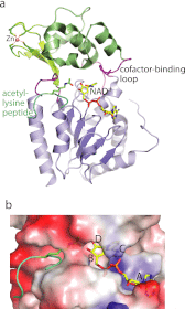
FIGURE 1.
FIGURE 1.
Overall sirtuin structure represented by Hst2. a, schematic of the overall structure of the protein bound to acetyllysine and carba-NAD+ substrates represented in stick model (Protein Data Bank code 1SZC). The small domain shown in green, the large domain in blue, and the four linking loops in purple. b, the substrate-binding cleft is shown in surface representation and is colored red for negative charge and blue for positive charge. The NAD+-binding region is divided into sites A–D.
FIGURE 2.
FIGURE 2.
Binding of sirtuin substrates. a–d, binding of NAD+. a, site A. Residues represented in blue stick form hydrogen bonds (dashed lines) with NAD+. The adenine base sits in a hydrophobic pocket shown in surface representation. yHst2, yeast Hst2; PDB, Protein Data Bank. b, sites B and C. NAD+ forms hydrogen bonds with Ile-117, Asp-118, and acetyllysine. Gly-32, Ala-33, Gly-34, Ile-41, and Phe-44 make van der Waals interactions with NAD+. Asn-116 forms a hydrogen bond with an ordered water molecule (w; blue sphere). c, sites B and C represented in the Sir2Tm-NAD+-acetyllysine complex. The nicotinamide ribose is rotated ∼90 °C compared with b. d, different conformations of the cofactor-binding loop. Hst2 bound to carba-NAD+ (purple) is superimposed on Hst2 bound to ADPR (blue), showing the different positions of Phe-44. e and f, binding of acetyllysine-containing p53 peptide bound to Sir2Tm. e, the acetyllysine peptide (light green) forms a β-staple with sirtuin residues (dark green). Only the backbone of the residues is shown except for where side chain interactions are made: Lys-381 (−1 position) forms hydrogen bonds with Asn-165 and Gly-163, and Met-384 (+2 position) makes van der Waals interactions with Phe-162 and Val-193. f, the acetyllysine side chain is buried in a hydrophobic tunnel and makes van der Waals interactions with sirtuin residues (blue stick). Val-160 also forms a hydrogen bond with the Nϵ of acetyllysine. g, succinyllysine (green) forms hydrogen bonds with SIRT5 residues (blue stick) and NAD+.
FIGURE 3.
FIGURE 3.
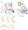
Inhibitor binding to sirtuins and NAD+ analog structures. a, nicotinamide (NAM) bound to the C pocket. Nicotinamide forms hydrogen bonds with Ile-102 and Asp-103 (blue stick; Sir2Af2) and π-stacking interactions with Phe-35 (purple). b, suramin (yellow sticks) bound to SIRT5. A dimer of SIRT5 binds to the symmetric suramin, and only one SIRT5 is shown for clarity. The succinyllysine-containing peptide and NAD+ (aqua lines; Protein Data Bank code 3RIY) are superimposed to show that suramin competes with both substrates for sirtuin binding. c, structures of NAD+ and its analogs in Corey-Pauling-Koltun colors. Yellow, carbon; dark yellow, sulfur; red, oxygen; blue, nitrogen; orange, phosphate. The AMP moiety is identical among the molecules and is not shown. The replacements from authentic molecules are indicated by red arrows.

References

    1. Bell S. D., Botting C. H., Wardleworth B. N., Jackson S. P., White M. F. (2002) The interaction of Alba, a conserved archaeal chromatin protein, with Sir2 and its regulation by acetylation. Science 296, 148–151 - PubMed
    1. Starai V. J., Celic I., Cole R. N., Boeke J. D., Escalante-Semerena J. C. (2002) Sir2-dependent activation of acetyl-CoA synthetase by deacetylation of active lysine. Science 298, 2390–2392 - PubMed
    1. Imai S., Guarente L. (2010) Ten years of NAD-dependent SIR2 family deacetylases: implications for metabolic diseases. Trends Pharmacol. Sci. 31, 212–220 - PMC - PubMed
    1. Kim E. J., Um S. J. (2008) SIRT1: roles in aging and cancer. BMB Rep. 41, 751–756 - PubMed
    1. Guarente L. (2007) Sirtuins in aging and disease. Cold Spring Harb. Symp. Quant. Biol. 72, 483–488 - PubMed

LinkOut - more resources