. 2012 Dec 7;287(50):41583-94.
doi: 10.1074/jbc.M112.415984. Epub 2012 Oct 20.

Human Rap1 interacts directly with telomeric DNA and regulates TRF2 localization at the telomere

Affiliations

Human Rap1 interacts directly with telomeric DNA and regulates TRF2 localization at the telomere

N Özlem Arat et al. J Biol Chem. .

Abstract

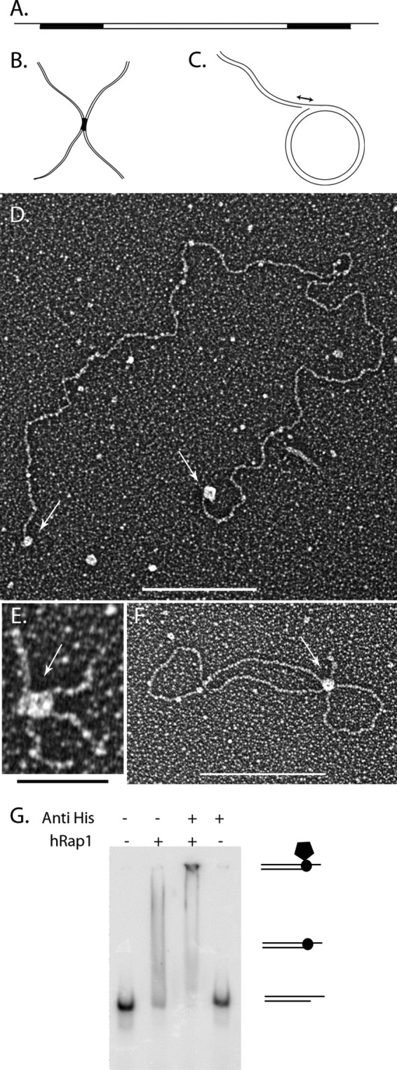
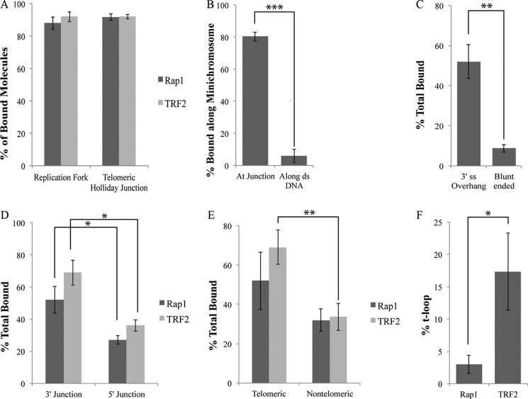
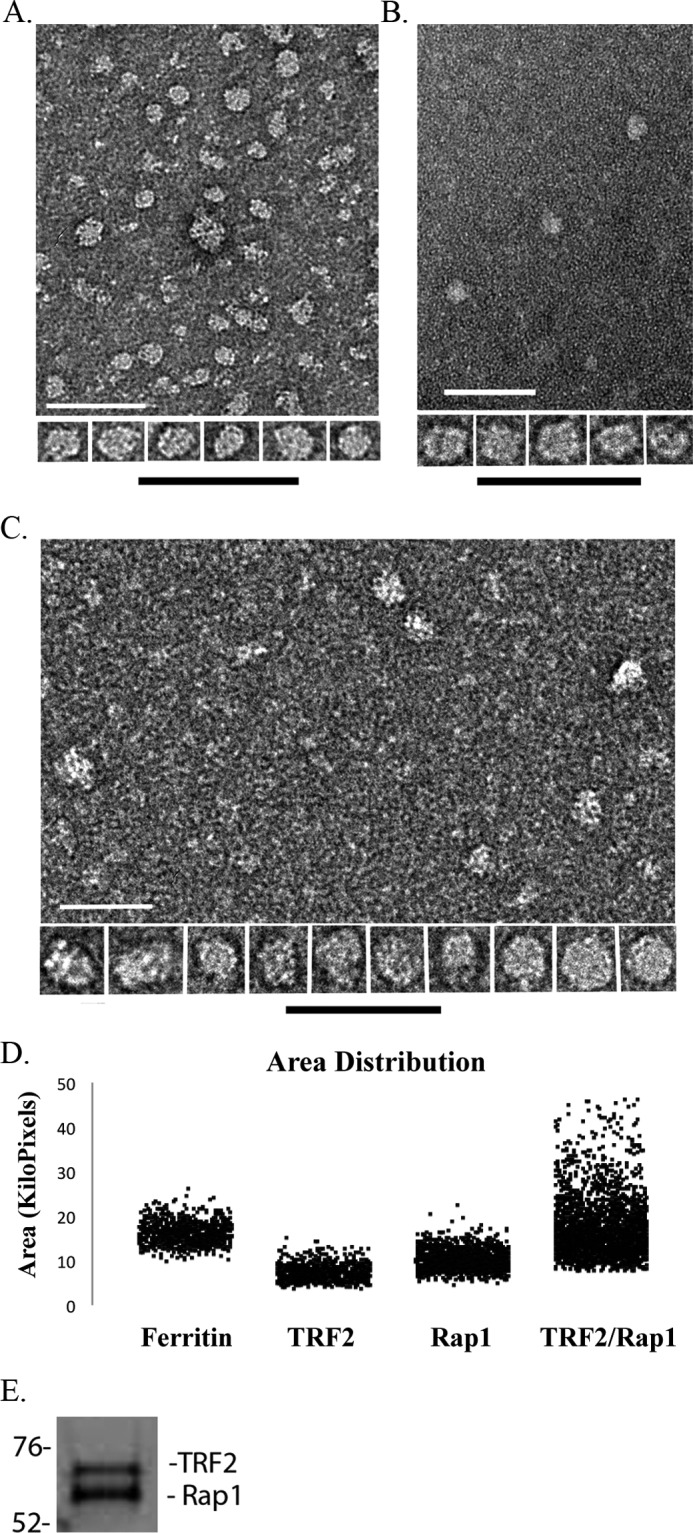
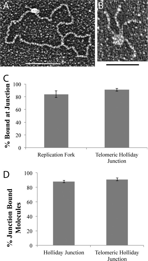
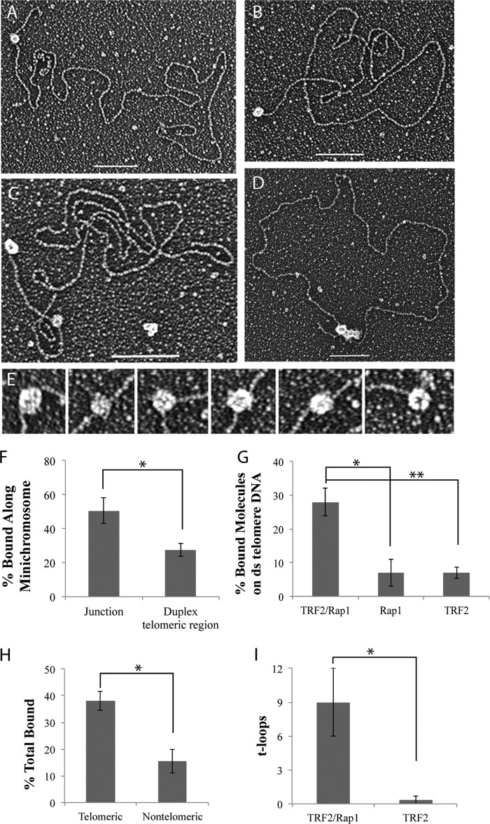
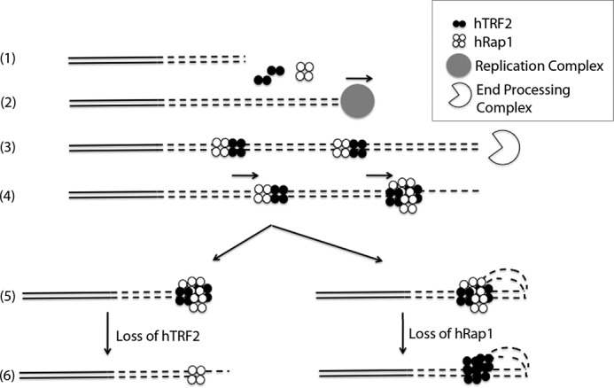
The TRF2-Rap1 complex suppresses non-homologous end joining and interacts with DNAPK-C to prevent end joining. We previously demonstrated that hTRF2 is a double strand telomere binding protein that forms t-loops in vitro and recognizes three- and four-way junctions independent of DNA sequence. How the DNA binding characteristics of hTRF2 to DNA is altered in the presence of hRap1 however is not known. Here we utilized EM and quantitative gel retardation to characterize the DNA binding properties of hRap1 and the TRF2-Rap1 complex. Both gel filtration chromatography and mass analysis from two-dimensional projections showed that the TRF2-Rap1 complex exists in solution and binds to DNA as a complex consisting of four monomers each of hRap1 and hTRF2. EM revealed for the first time that hRap1 binds to DNA templates in the absence of hTRF2 with a preference for double strand-single strand junctions in a sequence independent manner. When hTRF2 and hRap1 are in a complex, its affinity for ds telomeric sequences is 2-fold higher than TRF2 alone and more than 10-fold higher for telomeric 3' ends. This suggests that as hTRF2 recruits hRap1 to telomeric sequences, hRap1 alters the affinity of hTRF2 and its binding preference on telomeric DNA. Moreover, the TRF2-Rap1 complex has higher ability to re-model telomeric DNA than either component alone. This finding underlies the importance of complex formation between hRap1 and hTRF2 for telomere function and end protection.


Figures

FIGURE 1.
FIGURE 1.
hRap1 binds to model DNA templates. Schematic representations of the minichromosome (A), replication fork with a 25-nt gap (B), and Holliday junction templates (C). Black regions correspond to the telomeric DNA. Tungsten shadowcast images of hRap1 illustrate protein bound to the minichromosome (D), replication fork (E), and telomeric Holliday junction template (F). Data are shown in reverse contrast. Scale bar in D and F are 100 nm and is 50 nm in E. Shown is EMSA with 4% PAGE demonstrates hRap1 bound to the nontelomeric template with a 3′ overhang in the presence and absence of anti-His6 antibody (G).
FIGURE 2.
FIGURE 2.
hRap1 recognizes the 3′ ds-ss junction structures independent of sequence. A illustrates that both hRap1 and TRF2 have a strong and similar preference for binding to the ss-ds junction at replication fork with a 25-nt gap and the crossover at the HJ. B illustrates the strong preference for hRap1 binding to the end of the minichromosome containing a 3′ ss extension as contrasted to binding internally along the ds telomeric segment. C showed the strong preference for hRAP1 binding to the minichromosome containing a 3′ ss extension as contrasted to the same but blunt-ended DNA. D shows that both hRap1 and TRF2 prefer to bind at the ss-ds junction of DNAs with a 3′ overhang as contrasted to a 5′ overhang. E compares the binding to DNAs containing 3′ overhangs joined to either telomeric ds segments or non-telomeric. F compares the t-loop formation percentages of hRap1 and hTRF2 on the minichromosome. Each binding experiment was done in triplicate, and at least 100 molecules were counted. *, p < 0.05; **, p < 0.01; ***, p < 0.001.
FIGURE 3.
FIGURE 3.
Mass and oligomeric state analysis of hRap1, hTRF2, and the hRap1-TRF2 complex. Representative negative stained images of hTRF2 (A), hRap1 (B), and the TRF2-Rap1 complex (C) are shown as fields and an array of selected single particles at higher magnification. Bars are equivalent to 200 nm. Area distributions of single protein particles and ferritin as a size standard were calculated from two-dimensional projections of the EM images (D). The TRF2-Rap1 complex was separated using 10% SDS-PAGE and stained with Coomassie Orange to determine the ratio of hRap1 to hTRF2 (E).
FIGURE 4.
FIGURE 4.
The hTRF2-Rap1 complex recognizes replication forks and Holliday junctions. The TRF2-Rap1 complex localizes to the junction site of the replication fork (A) and the telomeric HJ DNA (B). Junction preference of the protein bound molecules on the DNA templates is in C, whereas D represents the sequence preference of the hTRF2-Rap1 complex on the HJ DNAs. Each EM binding reaction was done in triplicate, and 100 molecules each were counted. *, p < 0.05; **, p < 0.01; ***, p < 0.001.
FIGURE 5.
FIGURE 5.
The binding properties of the TRF2-Rap1 complex along the minichromosome. Tungsten shadowcast images of the TRF2-Rap1 complex on the minichromosome illustrate the protein bound to the ds-ss junction and/or to the ds telomeric DNA (A–C). An example of the TRF2-Rap1 complex bringing the ends of the minichromosome together to form a circle is depicted in D. Bars in A–D are equivalent to 100 nm. In high magnification images, the ds telomeric DNA appears to pass through the TRF2-Rap1 complex (E). Analysis of the binding preference of the TRF2-Rap1 complex along the minichromosome DNA is in F, showing significant binding to the internal duplex telomeric sequences. G further compares the binding percentages of hRap1, hTRF2, and the complex to the ds telomeric DNA segments of the minichromosomes. The effect of DNA sequence on the binding of the TRF2-Rap1 is shown in H. I demonstrates the comparison of the t-loops formed by the TRF2-Rap1 complex and hTRF2 at 10 nm protein concentration. Corresponding p values are shown by the different number of asterisks on each graph as described in the legend to Fig. 2.
FIGURE 6.
FIGURE 6.
Model for the TRF2-Rap1 complex loading onto and protecting the telomere. (1), the nontelomeric and the telomeric DNA are presented in solid lines and in dashed lines (2), respectively. The new telomeric DNA is synthesized in S phase (3). hTRF2 and hRap1 bind to the newly synthesized ds telomeric DNA as a complex and then end resection occurs forming the 3′ overhang (4). The TRF2-Rap1 complexes slide along the ds telomeric DNA toward the newly formed 3′ overhang (5). End protection by the TRF2-Rap1 complex occurs through t-loop formation or simply by blocking the exposed end (6). In the absence of hTRF2, hRap1 binds to the junction site to prevent NHEJ (or HDR in mice) and in the absence of hRap1, hTRF2 prevents DDR by forming t-loops.

Similar articles

Cited by

References

    1. Griffith J. D., Comeau L., Rosenfield S., Stansel R. M., Bianchi A., Moss H., de Lange T. (1999) Mammalian telomeres end in a large duplex loop. Cell 97, 503–514 - PubMed
    1. Liu D., O'Connor M. S., Qin J., Songyang Z. (2004) Telosome, a mammalian telomere-associated complex formed by multiple telomeric proteins. J. Biol. Chem. 279, 51338–51342 - PubMed
    1. Palm W., de Lange T. (2008) How shelterin protects mammalian telomeres. Annu. Rev. Genet. 42, 301–334 - PubMed
    1. Hsu H. L., Gilley D., Galande S. A., Hande M. P., Allen B., Kim S. H., Li G. C., Campisi J., Kohwi-Shigematsu T., Chen D. J. (2000) Ku acts in a unique way at the mammalian telomere to prevent end joining. Genes Dev. 14, 2807–2812 - PMC - PubMed
    1. Zhu X. D., Niedernhofer L., Kuster B., Mann M., Hoeijmakers J. H., de Lange T. (2003) ERCC1/XPF removes the 3′ overhang from uncapped telomeres and represses formation of telomeric DNA-containing double minute chromosomes. Mol. Cell 12, 1489–1498 - PubMed

Publication types

MeSH terms