. 2013 Jan 1;304(1):L70-81.
doi: 10.1152/ajplung.00342.2012. Epub 2012 Oct 19.

High pulsatility flow stimulates smooth muscle cell hypertrophy and contractile protein expression

Affiliations

High pulsatility flow stimulates smooth muscle cell hypertrophy and contractile protein expression

Devon Scott et al. Am J Physiol Lung Cell Mol Physiol. .

Retraction in

Abstract

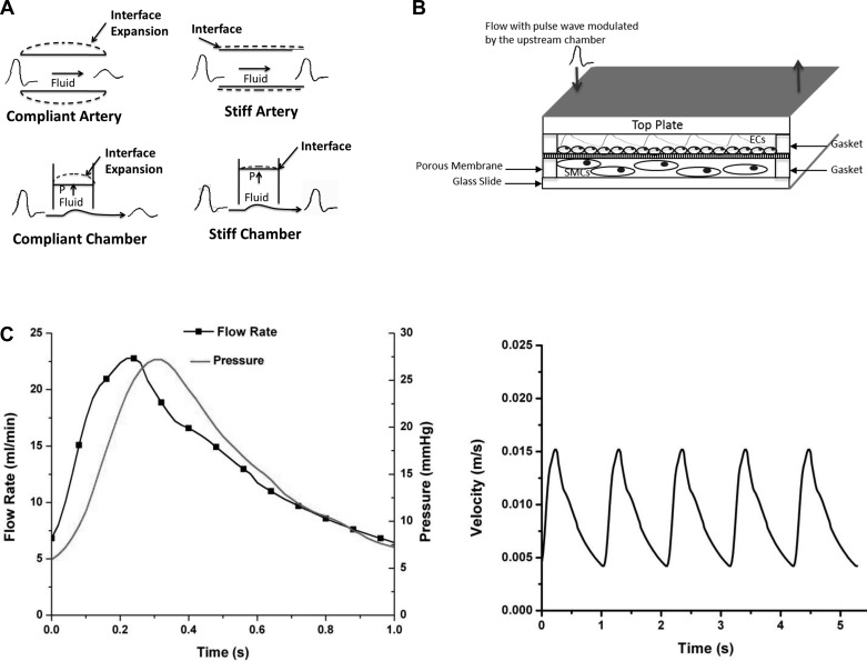
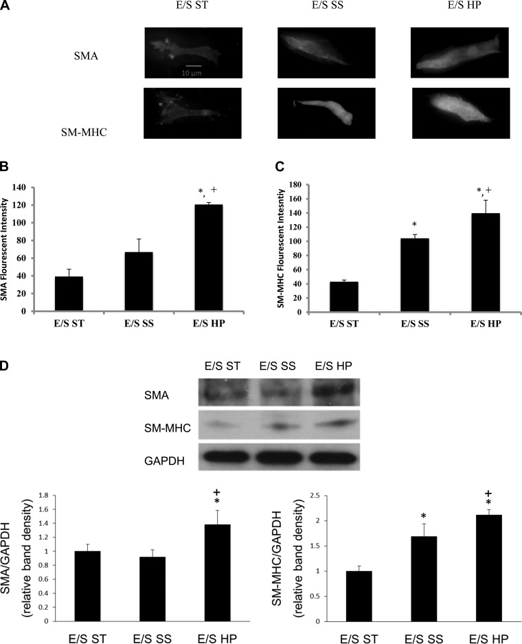
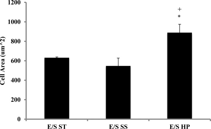
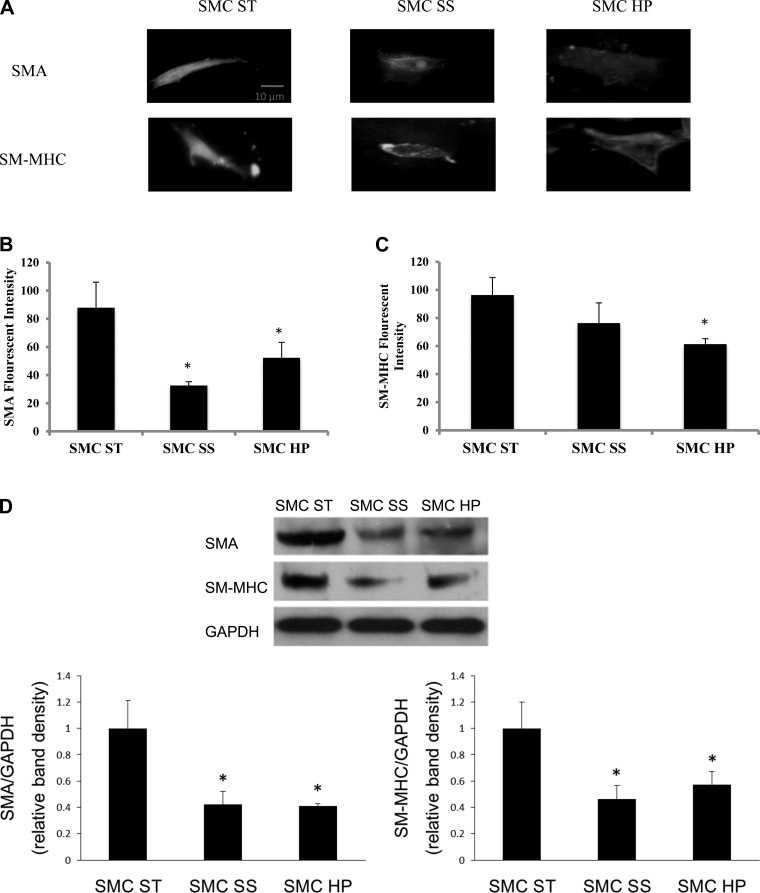
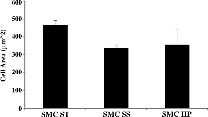
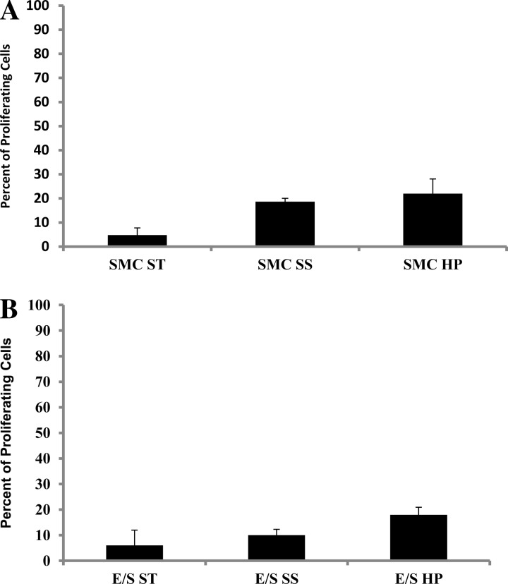
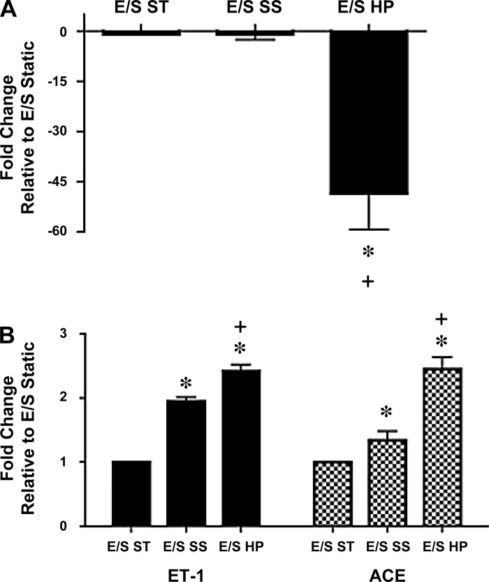
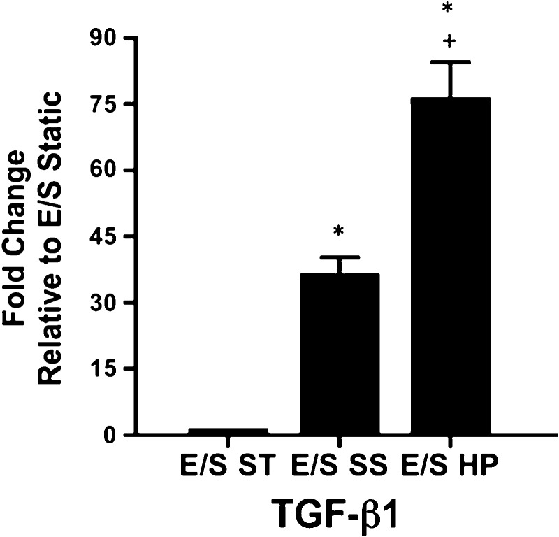
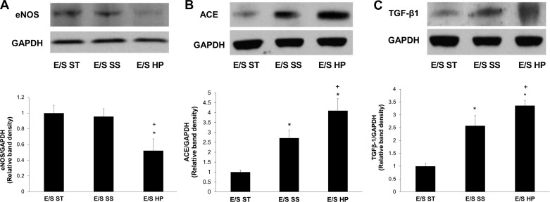
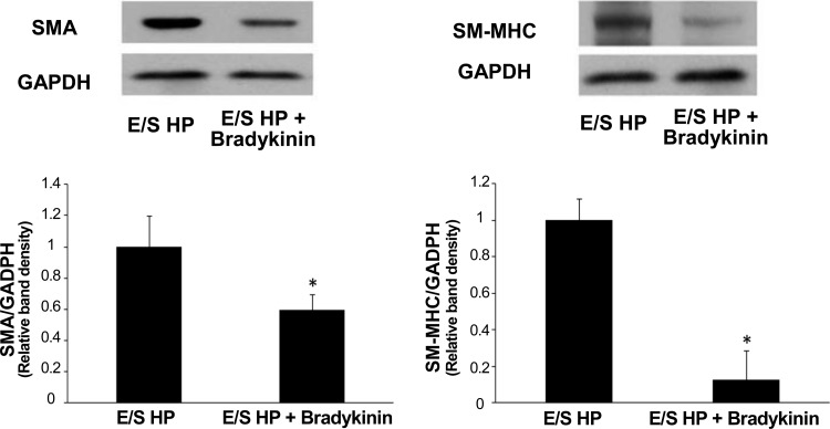
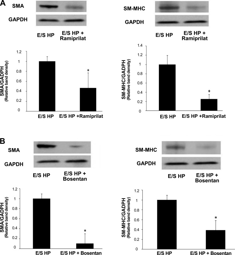
Proximal arterial stiffening is an important predictor of events in systemic and pulmonary hypertension, partly through its contribution to downstream vascular abnormalities. However, much remains undetermined regarding the mechanisms involved in the vascular changes induced by arterial stiffening. We therefore addressed the hypothesis that high pulsatility flow, caused by proximal arterial stiffening, induces downstream pulmonary artery endothelial cell (EC) dysfunction that in turn leads to phenotypic change of smooth muscle cells (SMCs). To test the hypothesis, we employed a model pulmonary circulation in which upstream compliance regulates the pulsatility of flow waves imposed onto a downstream vascular mimetic coculture composed of pulmonary ECs and SMCs. The effects of high pulsatility flow on SMCs were determined both in the presence and absence of ECs. In the presence of ECs, high pulsatility flow increased SMC size and expression of the contractile proteins, smooth muscle α-actin (SMA) and smooth muscle myosin heavy chain (SM-MHC), without affecting proliferation. In the absence of ECs, high pulsatility flow decreased SMC expression of SMA and SM-MHC, without affecting SMC size or proliferation. To identify the molecular signals involved in the EC-mediated SMC responses, mRNA and/or protein expression of vasoconstrictors [angiotensin-converting enzyme (ACE) and endothelin (ET)-1], vasodilator (eNOS), and growth factor (TGF-β1) in EC were examined. Results showed high pulsatility flow decreased eNOS and increased ACE, ET-1, and TGF-β1 expression. ACE inhibition with ramiprilat, ET-1 receptor inhibition with bosentan, and treatment with the vasodilator bradykinin prevented flow-induced, EC-dependent SMC changes. In conclusion, high pulsatility flow stimulated SMC hypertrophy and contractile protein expression by altering EC production of vasoactive mediators and cytokines, supporting the idea of a coupling between proximal vascular stiffening, flow pulsatility, and downstream vascular function.


Figures

Fig. 1.
Fig. 1.
Schematic illustrations of the flow coculture system. A: illustration of the concept of using a simplified engineering circulation to study the stiffening effects. A compliance proximal artery is able to buffer flow pulsatility from the heart while a stiff proximal artery is not able to expand to buffer these pulsations. The compliance-adjustment chamber mimics this function through a flow pressure-responsive interface modulated by the level of fluid in the chamber; the more fluid in the chamber the less pulse dampening, resulting in higher pulse flow (stiff artery), and the less fluid in the chamber the more flow dampening, resulting in low pulse flow (compliant artery). B: coculture flow chamber set up demonstrating the separation of endothelial cell (EC) monolayer and 3-dimensional smooth muscle cell (SMC) culture in collagen. The arrows demonstrate the flow direction. C: flow and pressure waves and the inlet velocity time history profiles for the high pulsatility flow used in this study.
Fig. 2.
Fig. 2.
High pulsatility flow increased SMC expression of contractile proteins in the presence of ECs. A: representative images of the SMCs stained with immunofluorescence for smooth muscle α-actin (SMA) and smooth muscle (SM)-myosin heavy chain (MHC), showing the effects of flow cocultures on SMCs. B: comparisons of SMA fluorescent intensities under static, steady, and high pulsatility flow coculture conditions. EC/SMC high pulsatility flow (E/S HP) showed increased SMA intensity. *P < 0.05 compared with EC/SMC static (E/S ST). +P < 0.05 compared with EC/SMC steady (E/S SS). C: comparisons of SM-MHC fluorescent intensities under static, steady, and high pulsatility flow coculture conditions. D: Western blot results showing similar changes of contractile proteins, SMA, and SM-MHC in SMC under static, steady, and high pulsatility flow cocultures.
Fig. 3.
Fig. 3.
High pulsatility flow increased SMC size in the presence of EC. The cell area of SMCs, as a measure of cell hypertrophy, increases under high pulsatility flow coculture (E/S HP) conditions compared with the steady flow (E/S SS) and static (E/S ST) conditions. *P < 0.05 compared with E/S ST. +P < 0.05 compared with E/S SS.
Fig. 4.
Fig. 4.
High pulsatility flow decreased SMC expression of contractile proteins in the absence of ECs compared with the static condition. A: representative images of the cells stained with immunofluorescence for SMA and SM-MHC showing the flow effects on SMCs with the absence of ECs. B: comparisons of SMA fluorescent intensities under static (SMC ST), steady (SMC SS), and high pulsatility (SMC HP) flow conditions. *P < 0.05 compared with SMC ST. C: comparisons of SM-MHC fluorescent intensities under static, steady, and high pulsatility flow conditions. D: Western blot results showing similar changes of contractile proteins, SMA and SM-MHC, in SMC under static, steady, and high pulsatility flow conditions.
Fig. 5.
Fig. 5.
High pulsatility flow did not change SMC size in the absence of ECs. The cell area of SMCs, as a measure of cell hypertrophy, did not significantly change under static (SMC ST), steady flow (SMC SS), and high pulsatility (SMC HP) flow conditions, without the presence of EC.
Fig. 6.
Fig. 6.
High pulsatility flow did not stimulate proliferation of quiescent SMCs. A: percent of proliferating SMC cultured without EC did not show statistically different increase in proliferation under high pulsatility flow (SMC HP) compared with the other conditions (SMC ST and SMC SS). B: percent of proliferating SMC cocultured with EC did not show statistically different increase under high pulsatility flow (E/S HP) compared with the other conditions (E/S ST and E/S SS).
Fig. 7.
Fig. 7.
High pulsatility flow altered EC production of vasoactive cytokines in the coculture conditions. The mRNA expressions for vasodilator [endothelial nitric oxide synthase (eNOS), A] as well as vasoconstrictors [endothelin (ET)-1 and angiotensin-converting enzyme (ACE), B] in different coculture conditions, including the static (E/S ST), steady flow (E/S SS), and high pulsatility flow (E/S HP) conditions. The mRNA levels under these conditions are normalized by the expressions from ECs without the presence of SMC in a static culture condition. *P < 0.05 compared with E/S ST. +P < 0.05 compared with E/S SS.
Fig. 8.
Fig. 8.
The mRNA expressions for transforming growth factor (TGF)-β1 in different coculture conditions, including the static (E/S ST), steady flow (E/S SS), and high pulsatility flow (E/S HP) conditions. The mRNA levels under these conditions are normalized by the expressions from ECs without the presence of SMC in a static culture condition. *P < 0.05 compared with E/S ST. +P < 0.05 compared with E/S SS.
Fig. 9.
Fig. 9.
Western blot results showed the protein expressions for eNOS (A), ACE (B), and TGF-β1 (C) in different coculture conditions, including the static (E/S ST), steady flow (E/S SS), and high pulsatility flow (E/S HP) flow conditions. The protein levels under these conditions are normalized by EC expressions of the housekeeping protein GAPDH. *P < 0.05 compared with E/S ST. +P < 0.05 compared with E/S SS.
Fig. 10.
Fig. 10.
Vasodilator bradykinin, which mediates nitric oxide production, reduced contractile protein expressions by SMCs cocultured with ECs under high pulsatility flow (E/S HP). Western blot results showed SMC expressions of SMA and SM-MHC under E/S HP were significantly reduced by bradykinin. *P < 0.05 compared with E/S HP.
Fig. 11.
Fig. 11.
Inhibitors of ACE or ET-1 receptor reduced contractile protein expressions by SMCs cocultured with ECs under high pulsatility flow (E/S HP). Western blot results showed SMC expressions of SMA and SM-MHC under E/S HP were significantly reduced by ACE inhibitor ramiprilat (A) and ET-1 receptor inhibitor bosentan (B). Both ramiprilat (10 μM) and bosentan (10 μM) were added in the flow media. *P < 0.05 compared with E/S HP.

References

    1. Adji A, O'Rourke MF, Namasivayam M. Arterial stiffness, its assessment, prognostic value, and implications for treatment. Am J Hypertens 24: 5–17, 2011 - PubMed
    1. Amann K, Gharehbaghi H, Stephen S, Mall G. Hypertrophy and hyperplasia of smooth muscle cells of small intramyocardial arteries in spontaneously hypertensive rats. Hypertension 25: 124–131, 1995 - PubMed
    1. Bakker EN, Matlung HL, Bonta P, de Vries CJ, van Rooijen N, Vanbavel E. Blood flow-dependent arterial remodelling is facilitated by inflammation but directed by vascular tone. Cardiovasc Res 78: 341–348, 2008 - PubMed
    1. Bjork JW, Tranquillo RT. Transmural flow bioreactor for vascular tissue engineering. Biotechnol Bioeng 104: 1197–1206, 2009 - PMC - PubMed
    1. Botney MD, Bahadori L, Gold LI. Vascular remodeling in primary pulmonary hypertension. Potential role for transforming growth factor-beta. Am J Pathol 144: 286–295, 1994 - PMC - PubMed

Publication types

MeSH terms

LinkOut - more resources