. 2012 Oct 19;338(6105):384-7.
doi: 10.1126/science.1226521.

Real-time evolution of new genes by innovation, amplification, and divergence

Affiliations

Real-time evolution of new genes by innovation, amplification, and divergence

Joakim Näsvall et al. Science. .

Abstract

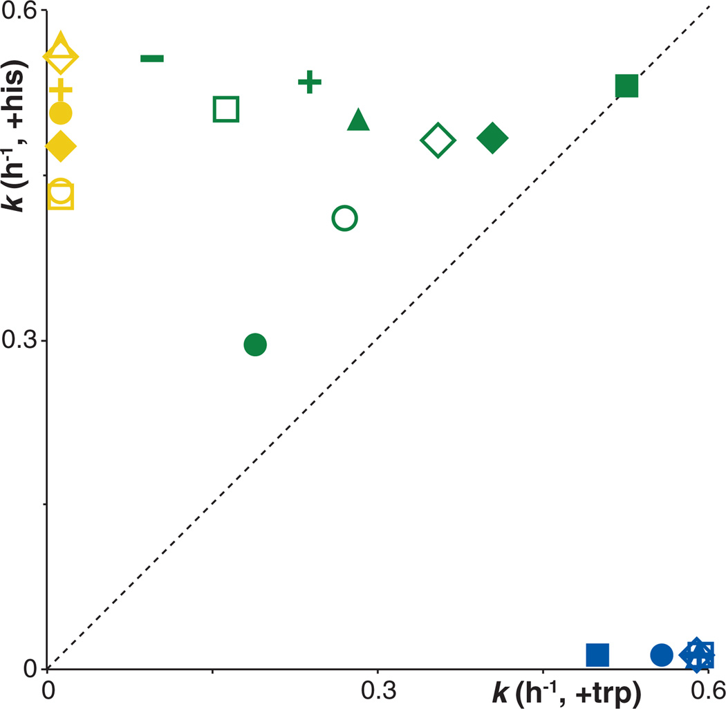
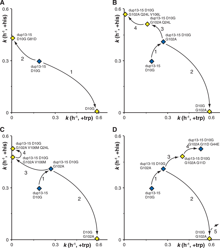
Gene duplications allow evolution of genes with new functions. Here, we describe the innovation-amplification-divergence (IAD) model in which the new function appears before duplication and functionally distinct new genes evolve under continuous selection. One example fitting this model is a preexisting parental gene in Salmonella enterica that has low levels of two distinct activities. This gene is amplified to a high copy number, and the amplified gene copies accumulate mutations that provide enzymatic specialization of different copies and faster growth. Selection maintains the initial amplification and beneficial mutant alleles but is relaxed for other less improved gene copies, allowing their loss. This rapid process, completed in fewer than 3000 generations, shows the efficacy of the IAD model and allows the study of gene evolution in real time.


Figures

Figure 1
Figure 1
(A) The IAD model. Innovation occurs when the ancestral gene (green) encodes a protein with the main function “A” and a minor activity “b.” Amplification occurs when an environmental change makes the b activity beneficial and selection favors variants with increased b activity. Divergence may occur in any one of the amplified gene copies that acquires a beneficial mutation that increases “B” activity (blue gene copy). After a B mutation, selection for the amplified array is relaxed, and segregation occurs to leave alleles with original A activity and the evolved B activity. (B) The isomerization reaction catalyzed by HisA and TrpF. The respective substrates and products differ in which chemical group (R) is attached to the 1′-amino group of the phosphoribosylamine. (C) Structure of the T-his element (linear insert) and its location on F′128 (circle) with the relative genetic elements on the F′ as shown (transposons; IS elements; replication origins; and the tra, lac, and mhp operons).
Figure 2
Figure 2
Trajectory for 3000 generations of evolution of the bifunctional parental gene (dup13-15, D10G) during selection for improved TrpF and HisA activities from one main parental lineage to the numerous variants found in daughter lineages. Mutations verified by sequencing are shown below the gene symbols. Red text indicates the identification of a new mutation for that lineage after the indicated number of generations. Additional lineages are shown in fig. S1, A to C. Asterisks next to a mutation indicate the presence of more than one subpopulation, differing in which of the indicated mutations they contain. Two asterisks indicate that only a subpopulation of the cells in the culture contained the indicated gene copy. A, Ala; Q, Gln; L, Leu; S, Ser; N, Asn; V, Val; M, Met; E, Glu; I, Ile.
Figure 3
Figure 3
Characteristics of 22 different evolved mutant gene variants. Each point represents the fitness of one specific mutant gene for its HisA activity on the x axis [assayed as growth rate (k) in minimal glycerol medium with added tryptophan] and TrpF activity on the y axis (assayed as growth rate in minimal glycerol medium with added histidine). Mutant genes fall into three main classes as indicated by the colors: Blue, HisA specialists [open diamond, hisA(wt); open triangle, D10G G102A; dash, D10G G102A V106M; cross, D10G; open square, D10G G102A S143N; solid circle, D10G R83C; solid square, D10G G102A V106M V88I]. Yellow, TrpF specialists (open triangle, dup13-15 D10G G102A Q24L V106L; open diamond, dup13-15 D10G G102A V106M V88I; cross, dup13-15 D10G G102A V106M Q24L; solid circle, dup13-15 D10G G102A Q24L V14:2M; solid diamond, dup13-15 D10G G102A V106M; open circle, dup13-15 D10G R83C; open square, dup13-15 D10G G81D). Green, generalist enzymes [solid circle, dup13-15 D10G (ancestral bifunctional gene); dash, dup13-15 D10G G102A V106M V45M; cross, dup13-15 D10G G102A Q24L G44E; solid square, dup13-15 D10G G102A G11D G44E; open square, dup13-15 D10G G102A Q24L; solid triangle, dup13-15 D10G G102A V88I; open diamond, dup13-15 D10G G102A S143N; solid diamond, dup13-15 D10G G102A G11D; open circle, dup13-15 D10G G102A].
Figure 4
Figure 4
Multiple evolutionary trajectories recovered through IAD. The x axis indicates the HisA activity (assayed as growth rate in minimal glycerol medium with added tryptophan); the y axis indicates the TrpF activity (assayed as growth rate in minimal glycerol medium with added histidine). (A) Evolution of specialist enzymes in which one activity is improved at the expense of the other. (B and C) Evolution of specialist enzymes after initial evolution of a generalist enzyme. (D) Evolution of a generalist enzyme with improvement of both activities. Arrows and numbers indicate the sequential order of appearance of the various mutations in the population. Yellow symbols denote gene variants that were always accompanied by another gene variant (generalist or with the complementary activity) in the same amplified array. Blue symbols denote gene variants that, at some point during the evolution, were the only variants found in the population.

Comment in

References

    1. Ochman H, Lawrence JG, Groisman EA. Nature. 2000;405:299. - PubMed
    1. Ohno S. Evolution by Gene Duplication. New York: Springer; 1970.
    1. Reams AB, Kofoid E, Savageau M, Roth JR. Genetics. 2010;184:1077. - PMC - PubMed
    1. Pettersson ME, Sun S, Andersson DI, Berg OG. Genetica. 2009;135:309. - PubMed
    1. Bergthorsson U, Andersson DI, Roth JR. Proc. Natl. Acad. Sci. U.S.A. 2007;104:17004. - PMC - PubMed

Publication types