. 2013 Jan 7;41(1):418-33.
doi: 10.1093/nar/gks965. Epub 2012 Oct 18.

Mitochondrial targeting of recombinant RNAs modulates the level of a heteroplasmic mutation in human mitochondrial DNA associated with Kearns Sayre Syndrome

Affiliations

Mitochondrial targeting of recombinant RNAs modulates the level of a heteroplasmic mutation in human mitochondrial DNA associated with Kearns Sayre Syndrome

Caroline Comte et al. Nucleic Acids Res. .

Abstract

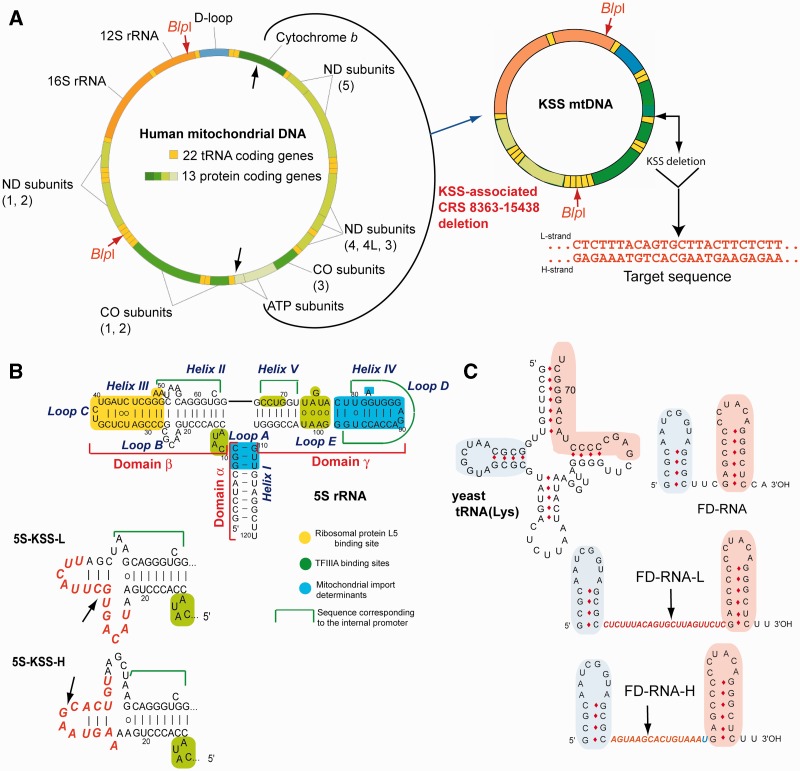
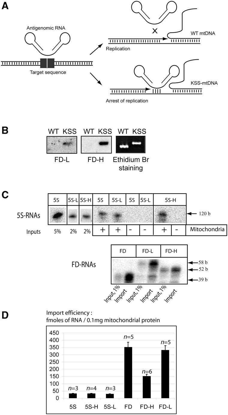
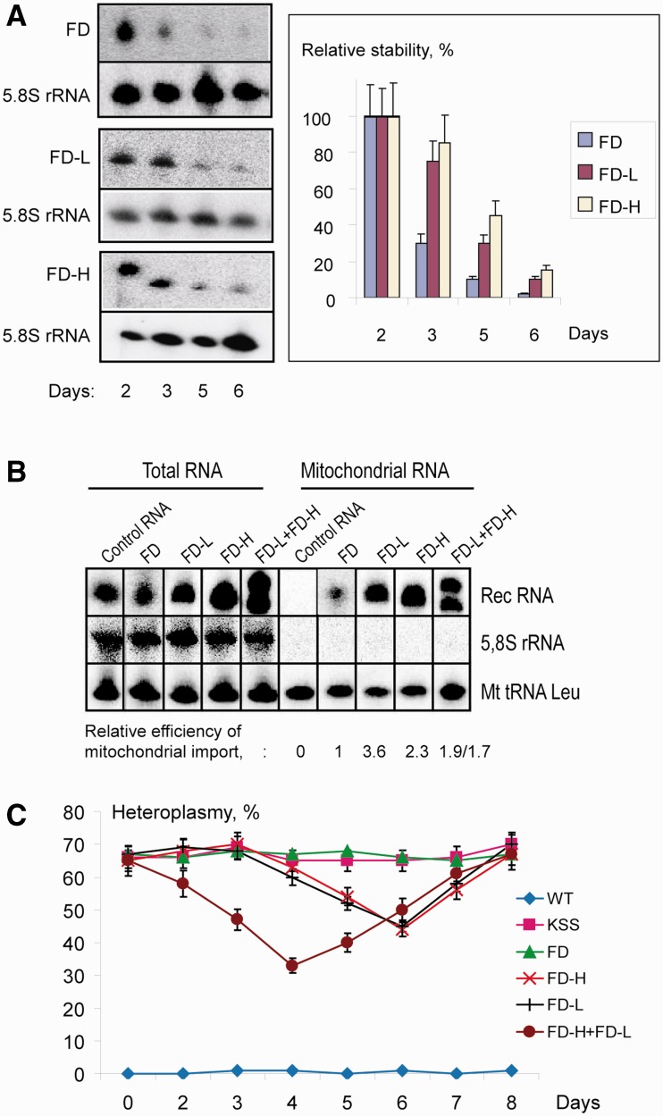
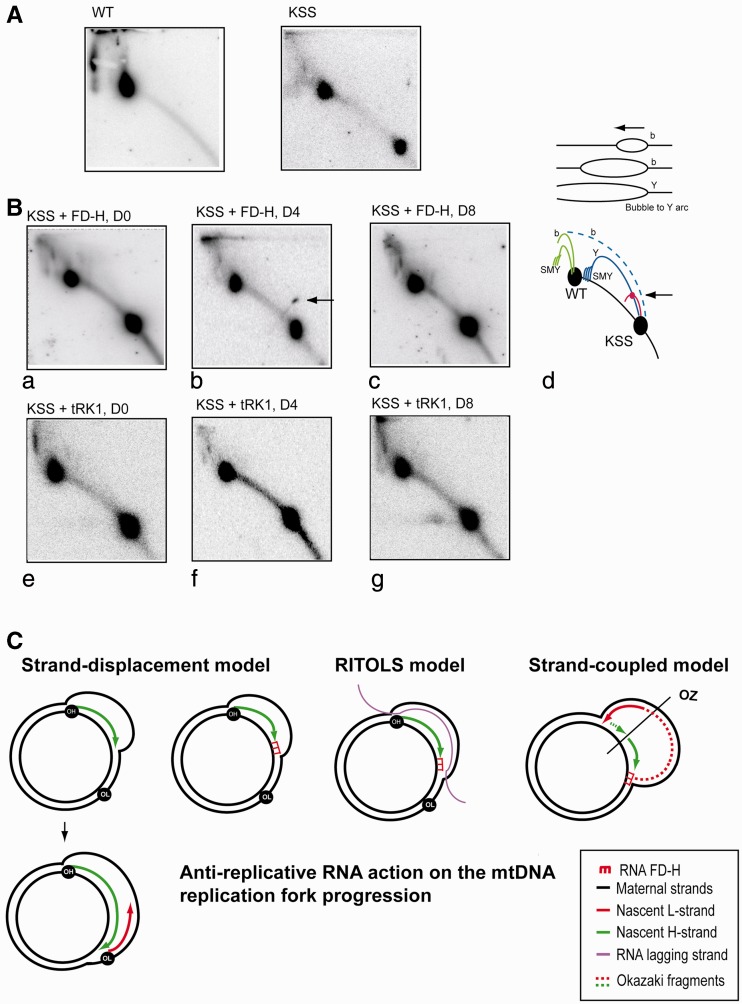
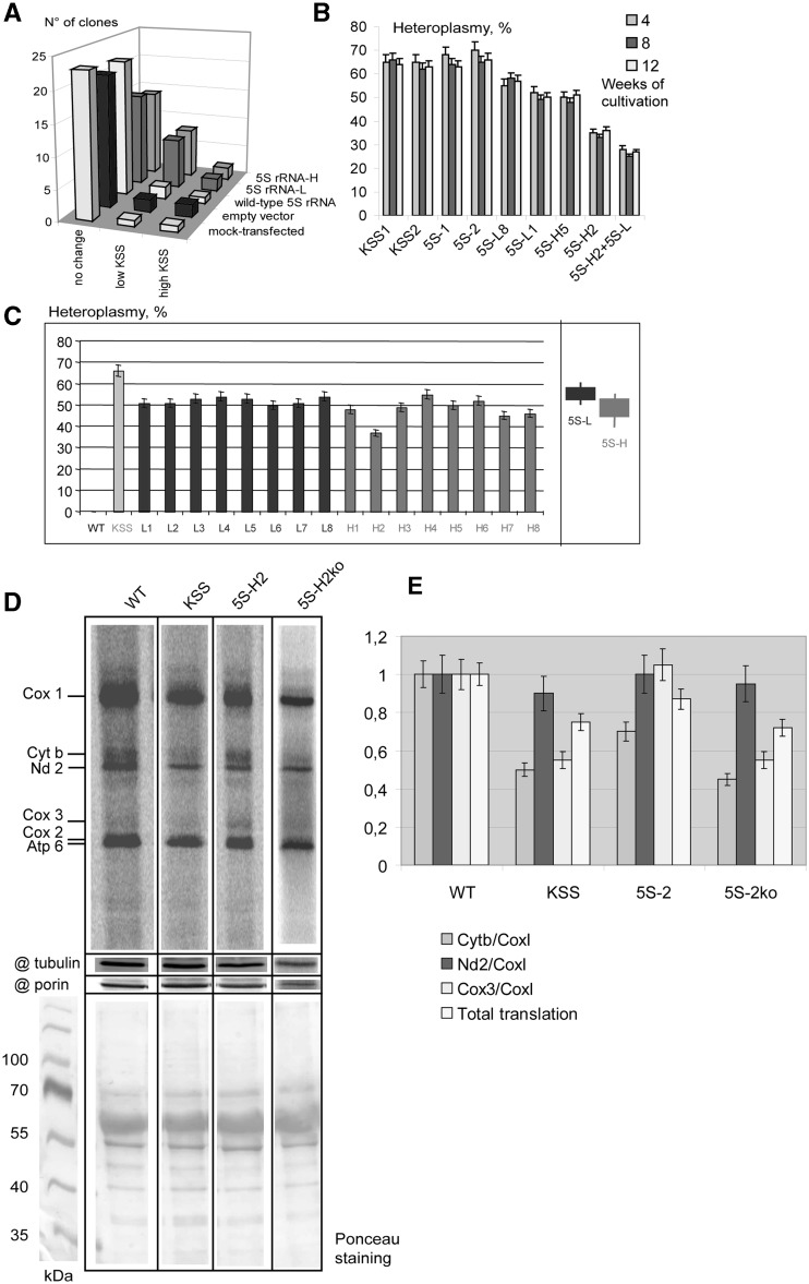
Mitochondrial mutations, an important cause of incurable human neuromuscular diseases, are mostly heteroplasmic: mutated mitochondrial DNA is present in cells simultaneously with wild-type genomes, the pathogenic threshold being generally >70% of mutant mtDNA. We studied whether heteroplasmy level could be decreased by specifically designed oligoribonucleotides, targeted into mitochondria by the pathway delivering RNA molecules in vivo. Using mitochondrially imported RNAs as vectors, we demonstrated that oligoribonucleotides complementary to mutant mtDNA region can specifically reduce the proportion of mtDNA bearing a large deletion associated with the Kearns Sayre Syndrome in cultured transmitochondrial cybrid cells. These findings may be relevant to developing of a new tool for therapy of mtDNA associated diseases.


Figures

Figure 1.
Figure 1.
KSS deletion in human mtDNA and anti-genomic RNAs design. (A) Genetic maps of wild-type and mutant KSS mtDNAs. Target sequence, the sequence of deletion boundaries is shown in red. (B) Secondary structure of human 5S rRNA and its functional elements. Below, secondary structures of recombinant 5S rRNA β-domains (inserts in red). The arrows show the deletion point. (C) The structures of yeast tRNALys, short synthetic FD-RNA and two recombinant RNAs containing inserts corresponding to L- and H-strands of KSS-mtDNA (in red). Two domains derived from the tRNA (32) are shown in pink and blue. The arrows show the deletion boundaries point.
Figure 2.
Figure 2.
(A) Scheme of the anti-genomic strategy. (B) Southern hybridization of wild-type or KSS mtDNA fragments with labeled recombinant RNAs (FD-L and FD-H) at 37°C. (C) In vitro import of the recombinant RNAs. Autoradiography of isolated from purified mitochondria RNA separated in denaturing 10% PAAG is presented; 5S rRNA derived molecules above and FD-RNA derived ones below. The size of RNA molecules is shown at the right. ‘Input’, 1–5% of RNA used for each assay, representing 30–150 fmoles of labeled RNA. Mitochondria (+) corresponds to the import assay, Mitochondria (−) to the mock import assay without mitochondria, used as a control for non-specific protein–RNA aggregation. (D) The import efficiencies, expressed in fmoles of protected RNA to 0.1 mg of mitochondrial protein, as described elsewhere (32); n, number of independent experiments; n = 3–6, as detailed upon each graph.
Figure 3.
Figure 3.
The effect of recombinant RNA mitochondrial targeting on heteroplasmy level in KSS cybrid cells. (A) Recombinant RNA stability in transiently transfected KSS cybrid cells. At the left, an example of Northern hybridization of total RNA isolated in different time after transfection (indicated below) with probes against FD-RNAs used for transfection (FD, FD-L or FD-H) and against 5.8S rRNA to quantify the level of recombinant RNA in the cells. At the right, time dependence of RNA decay, SD is calculated from at least four independent experiments. (B) Mitochondrial import of recombinant FD-RNAs in transiently transfected cybrid cells. Northern hybridizations of RNA extracted from cells or purified mitochondria 48 h after transfection. Above, RNAs used for transfection are indicated; control is an artificial RNA not imported into mitochondria (Supplementary Figure S5A). Relative efficiencies of mitochondrial import are shown below, import of FD-RNA is taken as 1 (±SD = 0.1, results of one from three independent experiments are presented). (C) Time dependence of KSS heteroplasmy level upon transient transfection of cybrid cells with various RNA (indicated at the right); KSS, mock-transfected cybrid cells. SD is calculated from three independent experiments.
Figure 4.
Figure 4.
Recombinant RNA affects mutant mtDNA replication in vivo. (A) The 2DNAGE electrophoresis analysis of mtDNA isolated from wild-type (WT) and KSS cybrid cells. MtDNA was digested with BlpI, blotted and hybridized with P32 labeled fragment of the Cyt b gene (Figure 1A). Radio autographs detect DNA fragment of 11 645bp for wild-type mtDNA and two fragments of 11 645 and 4570 bp for heteroplasmic mtDNA, indicated as WT and KSS on the cartoon at the panel d. (B) The 2DNAGE analysis of mtDNA isolated from KSS cybrid cells in different periods of time after transfection with RNA FD-H (panels a–c) and as a control with yeast tRNA (tRK1, panels e, f and g) immediately after transfection (panels a and e), in 4 days (panels b and f) and in 8 days (panels c and g). Panel d, schematic representation of replication intermediates: in green, replication intermediates corresponding to wild-type mtDNA and in blue, corresponding to mutant mtDNA. b, bubble arcs; Y, y arc; SMY, slow migrating y arcs. The site of presumptive replication pausing is shown by arrow. (C) Schematic representation of three mtDNA replication models (see ‘Discussion’ for details and references) and possible effect of anti-replicative recombinant RNA FD-H. OH, OL and OZ, origins of H-strand, L-strand, and strand-coupled replication, respectively.
Figure 5.
Figure 5.
Analysis of clones obtained after stable transfection of KSS cybrid cells with 5S rRNA genes. (A) Heteroplasmy variations in series of clones of KSS cybrid cells after stable transfection with 5S rRNA genes (indicated at the right). ‘No change,’ number of clones where the heteroplasmy shifts were comparable to natural fluctuation values (0–5%); ‘low KSS,’ the proportion of KSS mtDNA reduced by >10% and ‘high KSS,’ the proportion of KSS deletion increased by >10%. (B) Stability of the heteroplasmy level in individual clones. Two clones of each series are presented: KSS, mock-transfected KSS cybrid cells, clones from ‘no change’ set; 5S, cells transfected with the wild-type 5S rRNA gene, clones from ‘no change’ set are presented; 5S-H and 5S-L, cells transfected with corresponding recombinant 5S RNA genes, clones from ‘low KSS’ series, see also (C); 5S-H2+5S-L, clone 5S-H2 transfected with 5S-RNA-L gene. (C) Analysis of heteroplasmy levels in individual clones. All clones resulting from transfection with 5S-L and 5S-R RNAs where the hetroplasmy shift was >10% are presented, number of independent qPCR measurements n = 3. An example of such an assay, demonstrating that the shifts observed were significant and cannot be explained by errors of qPCR, is given in the Supplementary Figure S9. At the right, the summary of heteroplasmy shifts induced by 5S-L and 5S-H RNA is presented. (D) Analysis of mitochondrial protein synthesis in wild type, KSS cybrid cells, a clone of KSS cells expressing recombinant 5S rRNA (5S-H2) and the derivant of the same cell line after the loss of recombinant 5S rRNA expression (5S-H2ko), as indicated above the panel. Autoradiography of SDS-PAGE is demonstrated, products of mitochondrial translation are indicated at the left. @ tubulin and @ porin, immunoblotting of the same preparations, using as a loading control. The lower panel represents Ponceau S staining of the corresponding membrane. (E) The graphs represent the total level of mitochondrial translation (normalized to the amount of mitochondrial porin) and the ratio of signals corresponding to individual proteins. Cytb and Cox3, cytochrome b and subunit 3 of cytochrome oxidase, proteins encoded by the portion of mtDNA deleted in KSS mutants; CoxI and Nd2, subunit 1 of Cytochrom oxidaze and subunit 2 of NADH-dehyrogenase complexes, the both proteins encoded by the mtDNA region not touched by the KSS deletion.

References

    1. Ruiz-Pesini E, Lott MT, Procaccio V, Poole JC, Brandon MC, Mishmar D, Yi C, Kreuziger J, Baldi P, Wallace DC. An enhanced MITOMAP with a global mtDNA mutational phylogeny. Nucleic Acids Res. 2007;35:D823–D828. - PMC - PubMed
    1. Wallace DC. Mitochondrial diseases in man and mouse. Science. 1999;283:1482–1488. - PubMed
    1. Kolesnikova OA, Entelis NS, Mireau H, Fox TD, Martin RP, Tarassov IA. Suppression of mutations in mitochondrial DNA by tRNAs imported from the cytoplasm. Science. 2000;289:1931–1933. - PubMed
    1. Mahata B, Mukherjee S, Mishra S, Bandyopadhyay A, Adhya S. Functional delivery of a cytosolic tRNA into mutant mitochondria of human cells. Science. 2006;314:471–474. - PubMed
    1. Manfredi G, Fu J, Ojaimi J, Sadlock JE, Kwong JQ, Guy J, Schon EA. Rescue of a deficiency in ATP synthesis by transfer of MTATP6, a mitochondrial DNA-encoded gene, to the nucleus. Nat. Genet. 2002;30:394–399. - PubMed

Publication types