. 2013 Jan 7;41(1):264-76.
doi: 10.1093/nar/gks958. Epub 2012 Oct 18.

Possible steps of complete disassembly of post-termination complex by yeast eEF3 deduced from inhibition by translocation inhibitors

Affiliations

Possible steps of complete disassembly of post-termination complex by yeast eEF3 deduced from inhibition by translocation inhibitors

Shinya Kurata et al. Nucleic Acids Res. .

Abstract

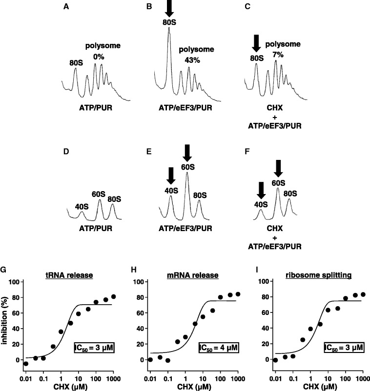
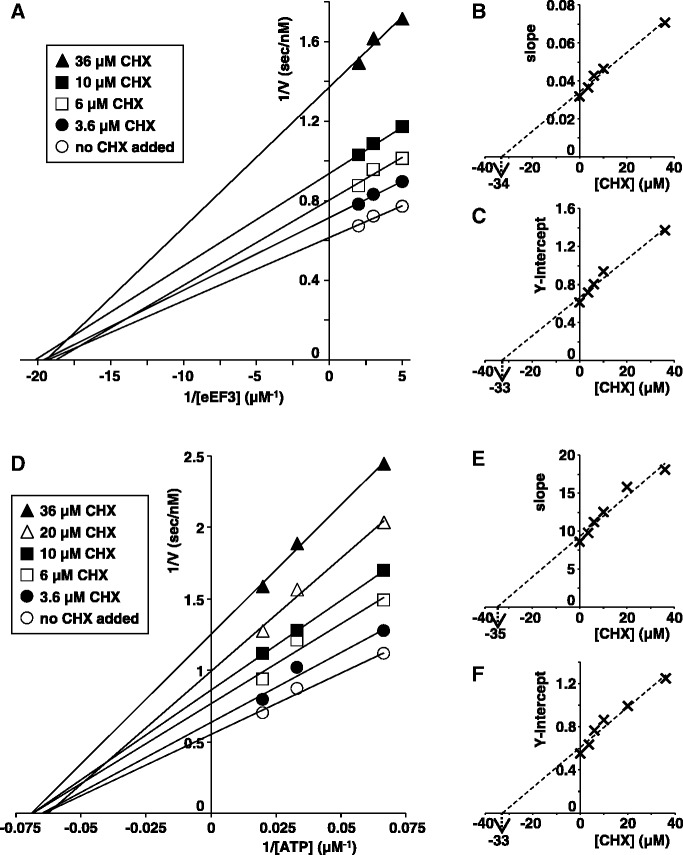
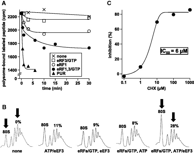
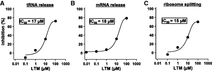
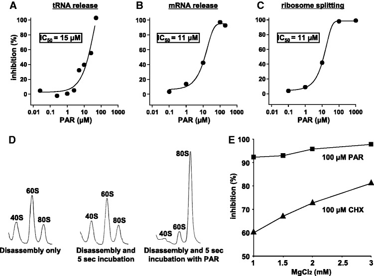
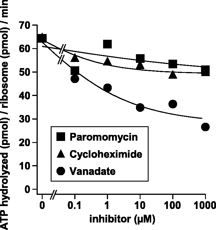
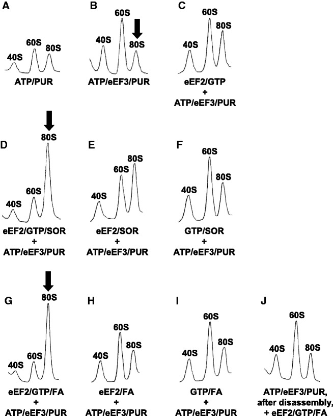
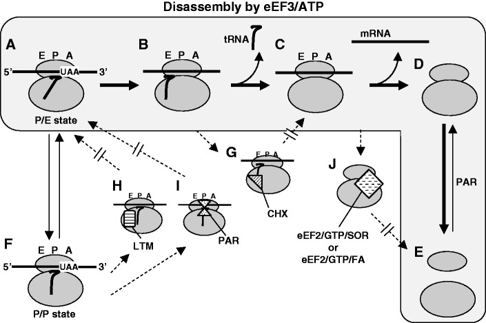
Ribosomes, after one round of translation, must be recycled so that the next round of translation can occur. Complete disassembly of post-termination ribosomal complex (PoTC) in yeast for the recycling consists of three reactions: release of tRNA, release of mRNA and splitting of ribosomes, catalyzed by eukaryotic elongation factor 3 (eEF3) and ATP. Here, we show that translocation inhibitors cycloheximide and lactimidomycin inhibited all three reactions. Cycloheximide is a non-competitive inhibitor of both eEF3 and ATP. The inhibition was observed regardless of the way PoTC was prepared with either release factors or puromycin. Paromomycin not only inhibited all three reactions but also re-associated yeast ribosomal subunits. On the other hand, sordarin or fusidic acid, when applied together with eEF2/GTP, specifically inhibited ribosome splitting without blocking of tRNA/mRNA release. From these inhibitor studies, we propose that, in accordance with eEF3's known function in elongation, the release of tRNA via exit site occurs first, then mRNA is released, followed by the splitting of ribosomes during the disassembly of post-termination complexes catalyzed by eEF3 and ATP.


Figures

Figure 1.
Figure 1.
Inhibitory effect of CHX on the disassembly of the PoTC by eEF3/ATP. (A–C) CHX inhibits the release of mRNA from PoTC. Sedimentation profiles of polysomes after the disassembly reaction of PoTC by eEF3/ATP under various conditions are shown. Final concentrations of each component are as follows: 50 µM ATP, 0.5 µM eEF3, 1 mM PUR, 360 µM CHX. The standard reactions for the disassembly of PoTC were stopped by buffer 25/150. This buffer converts the formed subunits to 80S ribosomes. The mixtures were subjected to SDGC with 150 000g for 75 min. (A) Only ATP/PUR was added. (B) Complete reaction mixture. (C) B in the presence of 360 µM CHX. Incubation was for 1 min at 30°C. The decrease of polysome area is accompanied by increase of the 80S ribosome (the arrows). Numbers on top of the polysome region indicate the percentage decrease of the area under each experimental condition. Note that in (C) (in the presence of CHX) the decrease of PoTC is significantly lower (43% versus 7%), and the increase of 80S ribosomes is less. (D–F) CHX inhibits the ribosome splitting of PoTC. For D–F, the reaction mixtures were fixed by formaldehyde to preserve subunits formed during the disassembly (10). They were centrifuged for 128 min at 150 000g to observe subunits. Remaining PoTC was pelleted and not observed under these sedimentation conditions. (D) Polysomes with ATP and PUR. (E) Complete reaction mixture. (F) E in the presence of 360 µM CHX. Note that subunits (arrows) were much less in F than E, indicating that the splitting of PoTC is inhibited by CHX. (G–I) Determination of IC50 of CHX on the three disassembly reactions of the PoTC by eEF3/ATP. Various amounts of CHX were added just before the disassembly reaction. IC50s of tRNA release (G), mRNA release (H) and the ribosome splitting (I) are shown.
Figure 2.
Figure 2.
CHX is a non-competitive inhibitor of both eEF3 and ATP. (A and D) Lineweaver–Burk plots of the disassembly reaction (mRNA release) inhibited by CHX at various concentrations of eEF3 (A) or ATP (D). (B and C) The slope (B) or Y intercept (C) of A was plotted against the concentrations of CHX. Ki and Ki’ of CHX were estimated to be 34 µM and 33 µM from B and C, respectively, where Ki and Ki’ are the dissociation constants for the enzyme–inhibitor (EI) complex or the enzyme–substrate–inhibitor (ESI) complex, respectively. (E and F) The slope (E) or Y intercept (F) of D was plotted against the concentrations of CHX. Ki and Ki’ of CHX were estimated to be 35 µM and 33 µM from E and F, respectively.
Figure 3.
Figure 3.
PoTC created by eRF1 and eRF3 is disassembled by eEF3. This reaction is also sensitive to CHX. (A) eRF1/eRF3/GTP releases 35S-labelled nascent peptides from the polysomes. The complete reaction mixture (60 µl) contained 20 pmol of labelled polysomes, 30 pmol of eRF1, 30 pmol of eRF3 and 20 nmol of GTP. Components of each reaction tube are as indicated in the inset. The reaction was carried out at 30°C and stopped by 1 mM anisomycin at the indicated times. The mixture was subjected to SDGC, and the radioactivity remaining on the polysomes was counted. (B) eEF3/ATP disassembles the PoTC formed by eRFs/GTP. Non-labelled polysomes were incubated with eRFs/GTP for 30 min to release peptide as in A. eEF3/ATP was then added, incubated for 3 min at 30°C to disassemble the formed PoTC. The reaction was stopped by 25 mM MgCl2, which also converts formed subunits to 80S ribosomes. The percentage of mRNA release was estimated from the reduction of the polysome and is indicated above the polysome region. Note the decrease (28% less) in polysome area and the increase of 80S peak between the extreme left and right panels (arrows). (C) CHX inhibits the disassembly of the PoTC formed by eRFs/GTP. Experimental conditions were as in B, except that various amounts of CHX were added just before the disassembly reaction, which was carried out for 1 min. Inhibitory effect of CHX (percent inhibition of mRNA release) is plotted against concentration of CHX.
Figure 4.
Figure 4.
Inhibitory effect of LTM on the disassembly of the PoTC by eEF3/ATP. The experimental conditions are the same as in Figure 1 except that various concentrations of LTM were added. Transfer RNA release (A) and mRNA release (B) from the PoTC and the ribosome splitting of the PoTC (C) are shown.
Figure 5.
Figure 5.
Dual effects of PAR. (A–C) Inhibition of PoTC disassembly. The experimental conditions are the same as in Figure 1 except that various concentrations of PAR were added. (D) Induction of subunit association by PAR. PoTC was disassembled into subunits by eEF3/ATP (left panel). The disassembled ribosomes were further incubated for 5 s at 30°C without PAR (middle panel) while the mixture shown in the right panel was incubated with 1 mM PAR for 5 s at 30°C. Then, the mixtures were subjected to SDGC, and sedimentation behaviours of the ribosomes are shown. (E) Effect of Mg2+ on the inhibitory effect of CHX and PAR. The disassembly of PoTC was carried out as in Figure 1, except that the reaction mixture contained 100 µM CHX (▴) or PAR (▪) and various concentrations of MgCl2 as indicated. The percent inhibition was calculated from the extent of the release of mRNA with antibiotic compared with that without antibiotic at each concentration of MgCl2.
Figure 6.
Figure 6.
Effects of CHX or PAR on ATP hydrolysis during the eEF3-catalysed disassembly of PoTC. The reaction mixture (15 µl) contained 0.1 µM PoTC, 0.5 µM eEF3, 50 µM [γ-32P]ATP, and various concentrations of inhibitors in buffer 3/150. Incubation was carried out at 30°C for 1 min, and the hydrolysis of ATP was plotted against the concentrations of inhibitors. Vanadate [an inhibitor of ATPase (32)] is a positive control and indicates the partial inhibition of the ATPase of eEF3/ribosome.
Figure 7.
Figure 7.
eEF2/GTP/SOR or eEF2/GTP/FA specifically inhibits eEF3/ATP-dependent splitting of 80S ribosomes of the PoTC. Sedimentation profiles of ribosomes after the disassembly reaction of PoTC by eEF3/ATP under various conditions are shown. Final concentrations of each component are as follows: 50 µM ATP, 0.5 µM eEF3, 1 mM PUR, 0.5 µM eEF2, 10 µM GTP, 100 µM SOR, 1 mM FA. An inhibitor mix (i.e. eEF2/GTP/SOR or eEF2/GTP/FA) was added before the disassembly reaction unless otherwise indicated. The reaction was stopped at 1 min by formaldehyde and centrifuged through 5–30% SDGC as in Figure 1D–F. (A) Only ATP/PUR was added. These ribosomal peaks represent background values. (B) Complete disassembly reaction. Note the increase of subunits. (C) Lack of effect of eEF2/GTP alone. (D) Complete reaction with inhibitor mix (eEF2/GTP/SOR). Note the increase of 80S peak and the decrease of subunits. (E) D without GTP. (F) D without eEF2. (G), (H) and (I) correspond to D, E and F, respectively, except that FA was added instead of SOR. Note the large peak of 80 S in D and G (arrows), presumably the complex with eEF2/inhibitors. (J) corresponds to G except that eEF2/GTP/FA was added after disassembly into subunits took place (1 min). Note that no increase of 80S ribosome was observed in this case.
Figure 8.
Figure 8.
eEF2/GTP/SOR or eEF2/GTP/FA inhibits ribosome splitting but not release of mRNA. (A–E) eEF3/ATP releases 80S ribosomes from the PoTC in the presence of eEF2/GTP/SOR or FA. (A) No additions. (B) ATP/PUR was added. (C) Complete reaction mix for disassembly. (D) Inhibitor mix (eEF2/GTP/SOR) was added to C. (E) Inhibitor mix (eEF2/GTP/FA) was added to C. Note the large peak of 80S in D and E (arrows), but the amount of polysomes are about the same for these three conditions. (F and G) Lack of incorporation of externally added labelled 60S subunits into 80S ribosomes formed from PoTC by eEF3/ATP in the presence of eEF2/GTP/SOR or FA. The reaction shown in Table 1 with 15 pmol of PoTC and other components was performed in the presence of 32P-labelled 60S subunits (0.25 pmol) with eEF2/GTP/SOR (F) or FA (G). The mixture was subjected to 5–30% SDGC, and both UV absorbance and radioactivity were measured for each fraction.
Figure 9.
Figure 9.
Proposed steps of the eEF3/ATP-dependent complete disassembly of PoTC. A and F are in equilibrium. A through E show the stepwise disassembly of PoTC (in the area coloured with pale grey). G through J show the presumed complexes of PoTC or partially disassembled PoTC with the inhibitors.

Similar articles

Cited by

References

    1. Hirokawa G, Demeshkina N, Iwakura N, Kaji H, Kaji A. The ribosome-recycling step: consensus or controversy? Trends Biochem. Sci. 2006;31:143–149. - PubMed
    1. Schneider-Poetsch T, Ju J, Eyler DE, Dang Y, Bhat S, Merrick WC, Green R, Shen B, Liu JO. Inhibition of eukaryotic translation elongation by cycloheximide and lactimidomycin. Nat. Chem. Biol. 2010;6:209–217. - PMC - PubMed
    1. Skogerson L, Wakatama E. A ribosome-dependent GTPase from yeast distinct from elongation factor 2. Proc. Natl Acad. Sci. USA. 1976;73:73–76. - PMC - PubMed
    1. Qin SL, Xie AG, Bonato MC, McLaughlin CS. Sequence analysis of the translational elongation factor 3 from Saccharomyces cerevisiae. J. Biol. Chem. 1990;265:1903–1912. - PubMed
    1. Triana-Alonso FJ, Chakraburtty K, Nierhaus KH. The elongation factor 3 unique in higher fungi and essential for protein biosynthesis is an E site factor. J. Biol. Chem. 1995;270:20473–20478. - PubMed

Publication types

MeSH terms