

Translocating the blood-brain barrier using electrostatics
- PMID: 23087614
- PMCID: PMC3468918
- DOI: 10.3389/fncel.2012.00044
Translocating the blood-brain barrier using electrostatics
Abstract
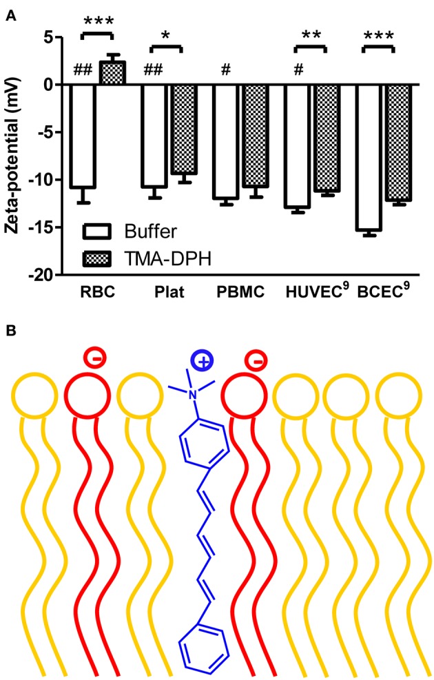
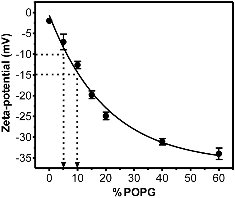
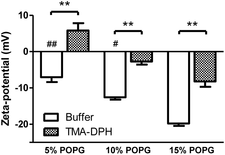
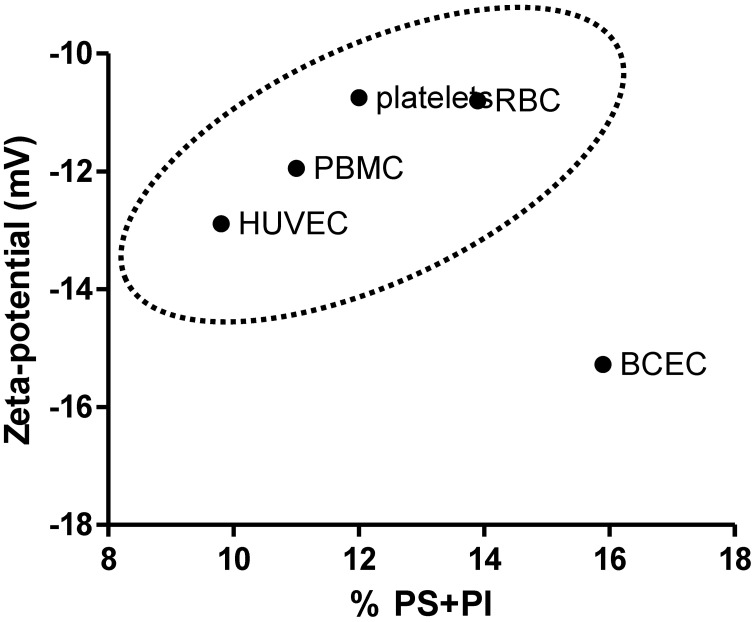
Mammalian cell membranes regulate homeostasis, protein activity, and cell signaling. The charge at the membrane surface has been correlated with these key events. Although mammalian cells are known to be slightly anionic, quantitative information on the membrane charge and the importance of electrostatic interactions in pharmacokinetics and pharmacodynamics remain elusive. Recently, we reported for the first time that brain endothelial cells (EC) are more negatively charged than human umbilical cord cells, using zeta-potential measurements by dynamic light scattering. Here, we hypothesize that anionicity is a key feature of the blood-brain barrier (BBB) and contributes to select which compounds cross into the brain. For the sake of comparison, we also studied the membrane surface charge of blood components-red blood cells (RBC), platelets, and peripheral blood mononuclear cells (PBMC). To further quantitatively correlate the negative zeta-potential values with membrane charge density, model membranes with different percentages of anionic lipids were also evaluated. From all the cells tested, brain cell membranes are the most anionic and those having their lipids mostly exposed, which explains why lipophilic cationic compounds are more prone to cross the blood-brain barrier.
Keywords: blood cells; blood-brain barrier; cell surface charge; drug targeting; zeta-potential.
Figures
References
-
- Anderson J. (1981). Concentration dependence of electrophoretic mobility. J. Colloid Interface Sci. 82, 248–250
-
- Buser C. A., Sigal C. T., Resh M. D., McLaughlin S. (1994). Membrane binding of myristylated peptides corresponding to the NH2 terminus of Src. Biochemistry 33, 13093–13101 - PubMed
-
- Cansell M., Gouygou J. P., Jozefonvicz J., Letourneur D. (1997). Lipid composition of cultured endothelial cells in relation to their growth. Lipids 32, 39–44 - PubMed
LinkOut - more resources
Full Text Sources
Other Literature Sources