. 2012 Jul 1;2(3):167-184.
doi: 10.4161/spmg.21800.

Meiosis in male Drosophila

Affiliations

Meiosis in male Drosophila

Bruce D McKee et al. Spermatogenesis. .

Abstract

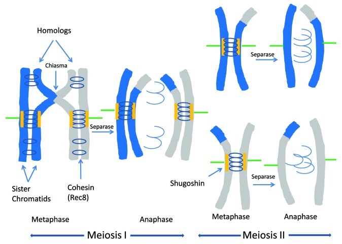
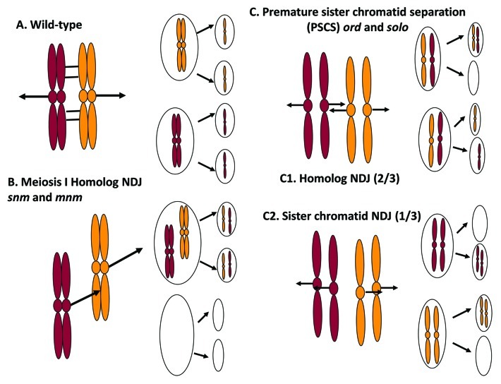
Meiosis entails sorting and separating both homologous and sister chromatids. The mechanisms for connecting sister chromatids and homologs during meiosis are highly conserved and include specialized forms of the cohesin complex and a tightly regulated homolog synapsis/recombination pathway designed to yield regular crossovers between homologous chromatids. Drosophila male meiosis is of special interest because it dispenses with large segments of the standard meiotic script, particularly recombination, synapsis and the associated structures. Instead, Drosophila relies on a unique protein complex composed of at least two novel proteins, SNM and MNM, to provide stable connections between homologs during meiosis I. Sister chromatid cohesion in Drosophila is mediated by cohesins, ring-shaped complexes that entrap sister chromatids. However, unlike other eukaryotes Drosophila does not rely on the highly conserved Rec8 cohesin in meiosis, but instead utilizes two novel cohesion proteins, ORD and SOLO, which interact with the SMC1/3 cohesin components in providing meiotic cohesion.


Figures

None
Figure 1. Meiotic chromosome segregation: how most eukaryotes do it. Homologous chromosomes segregate during meiosis I; sister chromatids segregate during meiosis II. In both divisions, chromosomes achieve bipolar orientation during prometaphase (not shown) and align at the spindle equator during metaphase. Sister chromatids are held together by cohesin complexes containing the meiosis-specific Rec8 subunit. In meiosis I, homologous chromosomes are linked by chiasmata at sites where homologous chromatids have undergone a crossover. Sister kinetochores attach to microtubules emanating from the same spindle pole. At anaphase I, Separase cleaves Rec8 cohesin only on chromosome arms, releasing chiasmata; Rec8 at centromeres is protected by Shugoshin. In meiosis II, the residual centromeric cohesion facilitates the attachment of sister kinetochores to microtubules emanating from opposite poles of the spindle. Sister chromatid separation at anaphase II is triggered by cleavage of the remaining Rec8 by Separase.
None
Figure 2. Meiosis I in Drosophila males. Meiosis occurs in cysts of 16 primary spermatocytes (only one shown). DNA replication is completed within 3 h of the last gonial mitosis and is followed by a lengthy growth period during which the three major chromosome pairs form separate territories by the start of stage S3 and remain separate throughout the rest of prophase I. Late in stage S6, the chromosomes condense to form four compact bivalents that achieve bipolar orientation (not shown) during prometaphase I and congress to the spindle equator at metaphase I. Euchromatic alleles are tightly paired during S1 and S2a but come unpaired, along with sister chromatids, at S2b/S3, coincident with territory establishment, and remain unpaired throughout the rest of meiosis I. The green dots represent fluorescent foci of GFP-LacI that accumulate at a specific chromosomal site where a 256mer array of lacO repeats is inserted. Pairing and cohesion status can be ascertained by counting spots, as shown. On chromosome diagrams, ovals represent centromeres and rectangles represent centromere-flanking heterochromatin.
None
Figure 3. Homolog pairing and localization of SNM, MNM and SOLO in wild-type spermatocytes. (A) Diagram of the X-Y pair showing restricted X-Y homology at shared rDNA loci. 359 is the 359 bp satellite sequence, a highly repeated element located between the centromere and the rDNA of the X chromosome and unique to the X. Circles represent centromeres, rectangles represent heterochromatin and line represents X euchromatin (not to scale). (B) FISH using fluorescently labeled probes specific for the 359 bp satellite (left panel) or the 240 bp repeats in the spacers of the rDNA repeats (right panel). DNA was counterstained with DAPI. Arrows point to X and Y chromosomes. 359 signal is clearly displaced from paired region in left panel, but 240 bp signal overlaps the paired region in the right panel. Note there is only one 240 bp signal signifying that the X and Y rDNA loci are paired. (C). Co-immuno-FISH analysis using 240 bp probe and anti-SNM antibody. DNA was counterstained with DAPI. After chromosome condensation and nucleolar dissolution, the 240 bp repeat and anti-SNM signals co-localize on the condensed X-Y pair until anaphase I. (D) MNM-GFP and SNM colocalize on the X-Y bivalent at prometaphase I. MNM-GFP detected by native fluorescence, SNM by an anti-SNM antibody and a FITC-conjugated secondary antibody. DNA was counterstained with DAPI. (E) MNM-GFP localizes to all four chromosome pairs throughout meiosis I (S4 and PMI shown). White arrows point to autosomal foci. X-Y signal is overexposed to allow autosomal signals to show. (F) SNM-Venus localizes to all four chromosome pairs throughout meiosis I (PMI shown). White arrows point to autosomal foci. DNA stained with DAPI. X-Y signal is overexposed in upper panel to allow autosomal signals to show. (G) Colocalization of Venus-SOLO and SMC1 during meiosis I. SMC1 was detected with an anti-SMC1 antibody and a FITC-conjugated secondary antibody, and Venus-SOLO was detected by native fluorescence. DNA was counterstained with DAPI. The spots of both proteins co-localize on centromeres throughout meiosis (based on colocalization with the centromere specific protein CID (not shown)).
None
Figure 4. Chromosome segregation patterns in Drosophila wild-type and meiotic mutants. (A) Wild-type bivalents are held together by the conjunction complex (cross-bars) which enables them to achieve bipolar orientation and segregate to opposite poles during meiosis I. Sister chromatids orient to the same pole (mono-orient) at meiosis I, then to opposite poles at meiosis II. (B) Homolog nondisjunction at meiosis I in snm and mnm mutants. Failure to maintain conjunction leads to premature homolog separation and random segregation at meiosis I. However, sister chromatids always mono-orient and segregate to the same pole at meiosis I. Meiosis II is normal and sister chromatids segregate to opposite poles. (C) Premature sister chromatid separation leads to random chromatid segregation in ord and solo mutants. Sister centromeres dissociate prematurely and orient randomly at meiosis I. The SNM-MNM conjunction complex is still present (not shown) and maintains bivalent integrity. In addition to the two types of balanced segregations pictured, unbalanced meiosis I segregations (3:1 or 4:0) can also occur but are mostly suppressed by the conjunction complex. (C1) Sister chromatids segregate to opposite poles at meiosis I (“equational” segregation) 2/3 of the time. Each secondary spermatocyte receives one chromatid from each homolog, which then segregate randomly. ¼ of the resulting spermatids will carry two homologous chromatids, resulting in homolog NDJ (same outcome as B but different mechanism). (C2) Sister chromatids segregate to the same pole (reductional segregation) at meiosis I 1/3 of time. Each secondary spermatocyte inherits a pair of sister chromatids (as in wild-type) but they are disconnected and segregate randomly at meiosis II. Diagram shows only the 2:0 segregations at meiosis II but 1:1 segregations are equally frequent. ¼ of the resulting spermatids will carry two sister chromatids, yielding sister chromatid NDJ.
None
Figure 5. Meiosis I chromosomes from wild-type, snm, solo and solo; snm spermatocytes at S5, prometaphase I (PMI) and metaphase I (MI). DNA was stained with DAPI and tubulin with an anti-tubulin antibody and detected with a FITC-conjugated secondary antibody. Note the poorly formed territories at S5 and the prematurely separated univalents at PMI and MI in snm spermatocytes. The bivalents appear normal in solo spermatocytes in this preparation but other squash methods reveal abnormal, loosely packed bivalents often with protruding single chromatids. solo; snm double mutants form multiple mini-territories at S5 which condense into single chromatids at PMI and MI.

Similar articles

Cited by

References

    1. Page SL, Hawley RS. Chromosome choreography: the meiotic ballet. Science. 2003;301:785–9. doi: 10.1126/science.1086605. - DOI - PubMed
    1. Petronczki M, Siomos MF, Nasmyth K. Un ménage à quatre: the molecular biology of chromosome segregation in meiosis. Cell. 2003;112:423–40. doi: 10.1016/S0092-8674(03)00083-7. - DOI - PubMed
    1. Hassold TJ, Hunt PA. To err (meiotically) is human: the genesis of human aneuploidy. Nat Rev Genet. 2001;2:280–91. doi: 10.1038/35066065. - DOI - PubMed
    1. Nicklas RB. How cells get the right chromosomes. Science. 1997;275:632–7. doi: 10.1126/science.275.5300.632. - DOI - PubMed
    1. Nasmyth K, Haering CH. Cohesin: its roles and mechanisms. Annu Rev Genet. 2009;43:525–58. doi: 10.1146/annurev-genet-102108-134233. - DOI - PubMed

LinkOut - more resources