. 2012 Jul 1;2(3):197-212.
doi: 10.4161/spmg.21798.

Drosophila spermiogenesis: Big things come from little packages

Affiliations

Drosophila spermiogenesis: Big things come from little packages

Lacramioara Fabian et al. Spermatogenesis. .

Abstract

Drosophila melanogaster spermatids undergo dramatic morphological changes as they differentiate from small round cells approximately 12 μm in diameter into highly polarized, 1.8 mm long, motile sperm capable of participating in fertilization. During spermiogenesis, syncytial cysts of 64 haploid spermatids undergo synchronous differentiation. Numerous changes occur at a subcellular level, including remodeling of existing organelles (mitochondria, nuclei), formation of new organelles (flagellar axonemes, acrosomes), polarization of elongating cysts and plasma membrane addition. At the end of spermatid morphogenesis, organelles, mitochondrial DNA and cytoplasmic components not needed in mature sperm are stripped away in a caspase-dependent process called individualization that results in formation of individual sperm. Here, we review the stages of Drosophila spermiogenesis and examine our current understanding of the cellular and molecular mechanisms involved in shaping male germ cell-specific organelles and forming mature, fertile sperm.


Figures

None
Figure 1. Diagrams illustrating stages of spermiogenesis (A-C) as well as nuclear shaping and chromatin condensation (D) in Drosophila. (A) In early round spermatids, mitochondria aggregate and fuse together to form the nebenkern, positioned near the haploid nucleus. The nebenkern consists of two mitochondrial derivatives wrapped around each other in a manner resembling an onion. The basal body, with a short axoneme surrounded by the ciliary sheath, embeds in the nuclear envelope. On the side of the nucleus opposite the basal body, Golgi bodies aggregate to form an acroblast and an acrosomal granule, from which will derive the acrosome. A dark, dense structure named the protein body forms in the nucleus, which is devoid of a nucleolus. Spermatids remain connected to each other via ring canals throughout most of spermiogenesis. (B) Each elongating spermatid contains an axoneme and two mitochondrial derivatives. Axoneme elongation occurs at the growing end, in the region ensheathed by the ciliary membrane. A ring of pericentriolar material called the centriolar adjunct forms around the basal body. Some of the proteins associated with the basal body, such as Unc, also localize to the ring centriole, a structure found at the edge of the ciliary cap that may be equivalent to a transition zone at the base of the cilium. Perinuclear microtubules organize at one side of the nucleus, forming the dense body. When the flagellar axoneme is about half its final length, the nucleus and the acrosome also start to elongate. (C) Each group of 64 spermatids is surrounded by two somatic cyst cells: a head cyst cell and a tail cyst cell. Elongated spermatid cysts are polarized, with all of the nuclei positioned in the head cyst cell, and the tails growing in the opposite direction. Fully elongated spermatids undergo individualization, a process in which F-actin containing investment cones form around the nuclei and then migrate in synchrony along the spermatids, stripping them of excess cytoplasm and unneeded organelles, and investing them with their own plasma membranes. As the cones move from head to tail, a cystic bulge forms around and in front of the cones. The excess cellular material is deposited in a waste bag at the end of the cyst. (D) As the nuclei elongate, they go through leaf, early canoe, late canoe and needle-shaped stages. The dense bodies and the acrosomes elongate together with the nuclei. Inside the nucleus, the chromatin is reorganized, and histones are replaced by transition proteins (Tpl) and then by protamines and Mst77F. During this histone to protamine transition, histones undergo various modifications (acetylation, ubiquitination), other proteins become sumoylated, transient breaks occur in the DNA strands and proteasome activity is high.
None
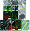
Figure 2. Micrographs illustrating selected stages of Drosophila spermiogenesis. (A) Round spermatid in onion stage with nucleus (arrowhead) and nebenkern (arrow). (B) Early elongated spermatids. Each nucleus contains a protein body (dark dot, arrowhead). (C) Mid elongated spermatid cyst. (D) Cross section of a mid elongated spermatid showing the axoneme (arrowhead), the major mitochondrial derivative starting to fill with paracrystalline material (arrow) and the minor mitochondrial derivative (red arrow). (E) Longitudinal section through a centriole near the cell periphery, showing the membrane cap (ciliary sheath, arrowhead). (F) Round spermatids (arrowhead) and late elongated cyst (arrow) showing basal bodies marked by GFP-PACT (green). (G) Round spermatids showing the ciliary membrane (arrowhead) contiguous with the plasma membrane marked by YFP-Skittles (green). (H) Early elongated spermatids showing the acroblast marked by Golph3-GFP (green). (I) Mature sperm nuclei showing the acrosome marked by GFP-Lamp (green). (J) Cross section through an onion stage spermatid. The nebenkern consists of two mitochondrial derivatives organized in concentric shells (arrowhead). The nucleus is only partially surrounded by a double membrane (arrow), where nuclear pores are located. (K) Longitudinal section showing a partially elongated nucleus, with basal body (arrowhead) and centriolar adjunct (red arrow) attached and with the dense body localized along one side of the nucleus (arrow). (L) Mid elongated cyst showing polarized spermatids, with nuclei at one end (arrow) and ring canals at the other (growing) end (arrowhead). Axonemes are marked by F-actin (phalloidin, red) and ring canals by anillin (green). (M) Mid elongated cyst showing polarized spermatids. Axonemes are marked by acetylated α-tubulin (green), which indicates stable microtubules. (N) Cross section through a cyst of individualized sperm, each with a nebenkern (black half circle, red arrow) and an axoneme (circle, red arrowhead). (O) Round spermatid nuclei. (P) Early elongated nuclei in leaf stage. (Q) Canoe stage nuclei. (R) Individualized nuclei. (S) Round nuclei showing dense bodies marked by anillin (green). (T) Canoe stage nuclei showing dense body marked by anillin (green). (U) Investment cones marked by F-actin (phalloidin, red). (V) Growing end of a late stage elongated spermatid cyst showing axonemes marked by acetylated α-tubulin (red) and the elongation complex marked by F-actin (phalloidin, green). (W) Elongated spermatid cysts in the individualization stage. Two cysts are at the beginning of individualization (arrowheads), whereas one cyst has just finished individualization and has a waste bag (arrow). Fully elongated axonemes are posttranslationally modified by polyglycylation (green). Investment cones are marked by F-actin (phalloidin, red). (X) Elongated cysts in various stages of individualization marked by activated Caspase 3 (green). The cystic bulge is visible in one of the cysts (arrowhead). (A-C) Phase-contrast images of live squashed preparations. (D, E, J, K, N) Transmission electron micrographs. (F, H) Merged phase-contrast and epifluorescence micrographs of live squashed preparations. (G, I, L, O, Q-T) Epifluorescence micrographs of fixed preparations. (M, P, U-V) Scanning confocal micrographs of fixed preparations. (W) Deconvolved confocal micrograph of fixed preparations. (X) Merged phase-contrast and epifluorescence micrograph of a fixed preparation. (F-I, L, M, S-T, W, X) Blue represents DNA stained with DAPI (in fixed preparations) or with Hoechst (in live squashed preparations). Scale bars = 1 μm in (J-K); = 5 μm in (A-B, F-I, L-M, O-X); = 20 μm in (C); = 200 nm in (D-E, N).

References

    1. Anderson WA. Cytodifferentiation of spermatozoa in Drosophila melanogaster: the effect of elevated temperature on spermiogenesis. Mol Gen Genet. 1967;99:257–73. doi: 10.1007/BF01797731. - DOI - PubMed
    1. Shoup JR. Spermiogenesis in wild type and in a male sterility mutant of Drosophila melanogaster. J Cell Biol. 1967;32:663–75. doi: 10.1083/jcb.32.3.663. - DOI - PMC - PubMed
    1. Tates AD. Cytodifferentiation during spermatogenesis in Drosophila melanogaster: An electron microscope study. Leiden: Rijksuniversiteit, 1971.
    1. Stanley HP, Bowman JT, Romrell LJ, Reed SC, Wilkinson RF. Fine structure of normal spermatid differentiation in Drosophila melanogaster. J Ultrastruct Res. 1972;41:433–66. doi: 10.1016/S0022-5320(72)90049-4. - DOI - PubMed
    1. Tokuyasu KT, Peacock WJ, Hardy RW. Dynamics of spermiogenesis in Drosophila melanogaster. I. Individualization process. Z Zellforsch Mikrosk Anat. 1972;124:479–506. doi: 10.1007/BF00335253. - DOI - PubMed

LinkOut - more resources