Growth conditions and environmental factors impact aerosolization but not virulence of Francisella tularensis infection in mice
- PMID: 23087911
- PMCID: PMC3468843
- DOI: 10.3389/fcimb.2012.00126
Growth conditions and environmental factors impact aerosolization but not virulence of Francisella tularensis infection in mice
Abstract
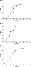
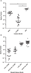
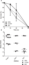
In refining methodology to develop a mouse model for inhalation of Francisella tularensis, it was noted that both relative humidity and growth media impacted the aerosol concentration of the live vaccine strain (LVS) of F. tularensis. A relative humidity of less than 55% had a negative impact on the spray factor, the ratio between the concentration of LVS in the aerosol and the nebulizer. The spray factor was significantly higher for LVS grown in brain heart infusion (BHI) broth than LVS grown in Mueller-Hinton broth (MHb) or Chamberlain's chemically defined medium (CCDM). The variability between aerosol exposures was also considerably less with BHI. LVS grown in BHI survived desiccation far longer than MHb-grown or CCDM-grown LVS (~70% at 20 min for BHI compared to <50% for MHb and CCDM). Removal of the capsule by hypertonic treatment impacted the spray factor for CCDM-grown LVS or MHb-grown LVS but not BHI-grown LVS, suggesting the choice of culture media altered the adherence of the capsule to the cell membrane. The choice of growth media did not impact the LD(50) of LVS but the LD(99) of BHI-grown LVS was 1 log lower than that for MHb-grown LVS or CCDM-grown LVS. Splenomegaly was prominent in mice that succumbed to MHb- and BHI-grown LVS but not CCDM-grown LVS. Environmental factors and growth conditions should be evaluated when developing new animal models for aerosol infection, particularly for vegetative bacterial pathogens.
Keywords: Francisella tularensis; aerosol exposure; mice; respiratory infection; tularemia.
Figures


Similar articles
-
Adaptation of Francisella tularensis to the mammalian environment is governed by cues which can be mimicked in vitro.Infect Immun. 2008 Oct;76(10):4479-88. doi: 10.1128/IAI.00610-08. Epub 2008 Jul 21. Infect Immun. 2008. PMID: 18644878 Free PMC article.
-
Development, Characterization, and Standardization of a Nose-Only Inhalation Exposure System for Exposure of Rabbits to Small-Particle Aerosols Containing Francisella tularensis.Infect Immun. 2019 Jul 23;87(8):e00198-19. doi: 10.1128/IAI.00198-19. Print 2019 Aug. Infect Immun. 2019. PMID: 31085702 Free PMC article.
-
Differential Growth of Francisella tularensis, Which Alters Expression of Virulence Factors, Dominant Antigens, and Surface-Carbohydrate Synthases, Governs the Apparent Virulence of Ft SchuS4 to Immunized Animals.Front Microbiol. 2017 Jun 22;8:1158. doi: 10.3389/fmicb.2017.01158. eCollection 2017. Front Microbiol. 2017. PMID: 28690600 Free PMC article.
-
A Francisella tularensis live vaccine strain (LVS) mutant with a deletion in capB, encoding a putative capsular biosynthesis protein, is significantly more attenuated than LVS yet induces potent protective immunity in mice against F. tularensis challenge.Infect Immun. 2010 Oct;78(10):4341-55. doi: 10.1128/IAI.00192-10. Epub 2010 Jul 19. Infect Immun. 2010. PMID: 20643859 Free PMC article.
-
Adherence and uptake of Francisella into host cells.Virulence. 2013 Nov 15;4(8):826-32. doi: 10.4161/viru.25629. Epub 2013 Jul 10. Virulence. 2013. PMID: 23921460 Free PMC article. Review.
Cited by
-
A Vibrating Mesh Nebulizer as an Alternative to the Collison Three-Jet Nebulizer for Infectious Disease Aerobiology.Appl Environ Microbiol. 2019 Aug 14;85(17):e00747-19. doi: 10.1128/AEM.00747-19. Print 2019 Sep 1. Appl Environ Microbiol. 2019. PMID: 31253680 Free PMC article.
-
Cross Sectional Study and Risk Factors Analysis of Francisella tularensis in Soil Samples in Punjab Province of Pakistan.Front Cell Infect Microbiol. 2019 Apr 5;9:89. doi: 10.3389/fcimb.2019.00089. eCollection 2019. Front Cell Infect Microbiol. 2019. PMID: 31024860 Free PMC article.
-
Development of an aerosol model of Cryptococcus reveals humidity as an important factor affecting the viability of Cryptococcus during aerosolization.PLoS One. 2013 Jul 23;8(7):e69804. doi: 10.1371/journal.pone.0069804. Print 2013. PLoS One. 2013. PMID: 23894542 Free PMC article.
-
Differential In Vitro Cultivation of Francisella tularensis Influences Live Vaccine Protective Efficacy by Altering the Immune Response.Front Immunol. 2018 Jul 10;9:1594. doi: 10.3389/fimmu.2018.01594. eCollection 2018. Front Immunol. 2018. PMID: 30042767 Free PMC article.
-
Differences in aerosolization of Rift Valley fever virus resulting from choice of inhalation exposure chamber: implications for animal challenge studies.Pathog Dis. 2014 Jul;71(2):227-33. doi: 10.1111/2049-632X.12157. Epub 2014 Mar 13. Pathog Dis. 2014. PMID: 24532259 Free PMC article.
References
-
- (2002). New drug and biological drug products; evidence needed to demonstrate effectiveness of new drugs when human efficacy studies are not ethical or feasible. Final rule. Fed. Regist. 67, 37988–37998 - PubMed
-
- FDA. (2009). Guidance for Industry: Animal Models–Essential Elements to Address Efficacy Under the Animal Rule. Available online at: http://www.fda.gov/downloads/Drugs/GuidanceComplianceRegulatoryInformati...(Accessed).
-
- Bandara A. B., Champion A. E., Wang X., Berg G., Apicella M. A., McLendon M., et al. (2011). Isolation and mutagenesis of a capsule-like complex (CLC) from Francisella tularensis, and contribution of the CLC to F. tularensis virulence in mice. PLoS ONE 6:e19003 10.1371/journal.pone.0019003 - DOI - PMC - PubMed
-
- Brasel T., Agans K., Barr E., Gonzales V., Romero F., Storch S., et al. (2009). Comparison of Francisella tularensis LVS and SCHU S4 bioaerosols, in Poster Presented at: 2009 ASM Biodefense and Emerging Infectious Diseases Meeting (Baltimore, MD: ).
Publication types
MeSH terms
Substances
LinkOut - more resources
Full Text Sources
Research Materials

