. 2013 May 1;18(13):1537-48.
doi: 10.1089/ars.2012.4666. Epub 2012 Dec 10.

NADPH oxidase-mediated triggering of inflammasome activation in mouse podocytes and glomeruli during hyperhomocysteinemia

Affiliations

NADPH oxidase-mediated triggering of inflammasome activation in mouse podocytes and glomeruli during hyperhomocysteinemia

Justine M Abais et al. Antioxid Redox Signal. .

Abstract

Aim: Our previous studies have shown that NOD-like receptor protein (NALP3) inflammasome activation is importantly involved in podocyte dysfunction and glomerular sclerosis induced by hyperhomocysteinemia (hHcys). The present study was designed to test whether nicotinamide adenine dinucleotide phosphate (NADPH) oxidase-mediated redox signaling contributes to homocysteine (Hcys)-induced activation of NALP3 inflammasomes, an intracellular inflammatory machinery in podocytes in vitro and in vivo.

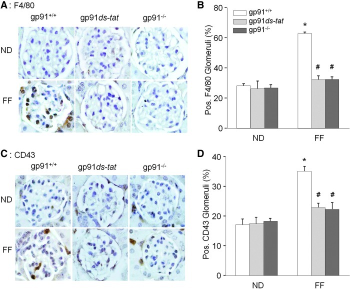
Results: In vitro confocal microscopy and size-exclusion chromatography revealed that upon NADPH oxidase inhibition by gp91(phox) siRNA, gp91ds-tat peptide, diphenyleneiodonium, or apocynin, aggregation of inflammasome proteins NALP3, apoptosis-associated speck-like protein (ASC), and caspase-1 was significantly attenuated in mouse podocytes. This NADPH oxidase inhibition also resulted in diminished Hcys-induced inflammasome activation, evidenced by reduced caspase-1 activity and interleukin-1β production. Similar findings were observed in vivo where gp91(phox-/-) mice and mice receiving a gp91ds-tat treatment exhibited markedly reduced inflammasome formation and activation. Further, in vivo NADPH oxidase inhibition protected the glomeruli and podocytes from hHcys-induced injury as shown by attenuated proteinuria, albuminuria, and glomerular sclerotic changes. This might be attributed to the fact that gp91(phox-/-) and gp91ds-tat-treated mice had abolished infiltration of macrophages and T-cells into the glomeruli during hHcys.

Innovation: Our study for the first time links NADPH oxidase to the formation and activation of NALP3 inflammasomes in podocytes.

Conclusion: Hcys-induced NADPH oxidase activation is importantly involved in the switching on of NALP3 inflammasomes within podocytes, which leads to the downstream recruitment of immune cells, ultimately resulting in glomerular injury and sclerosis.


Figures

FIG. 1.
FIG. 1.
NADPH oxidase inhibition attenuates inflammasome formation induced by hHcys in podocytes. (A) Confocal images representing the colocalization of NALP3 (green) with ASC (red) and NALP3 (green) with caspase-1 (red) in podocytes. (B) Summarized data showing the fold change of Pearson correlation coefficient (PCC) for the colocalization of NALP3 with ASC and NALP3 with caspase-1. (n=6–7). Ctrl, Control; Vehl, Vehicle; gp91pep, gp91ds-tat; DPI, diphenyleneiodonium; Scram, Scramble siRNA; ASCsi, ASC siRNA; gp91si, gp91phox siRNA. *P<0.05 vs. Control; #P<0.05 vs. Hcys. NADPH, nicotinamide adenine dinucleotide phosphate; Hcys, homocysteine; NALP3, NOD-like receptor protein; ASC, apoptosis-associated speck-like protein. (To see this illustration in color, the reader is referred to the web version of this article at www.liebertpub.com/ars.)
FIG. 2.
FIG. 2.
Distribution of inflammasome components after size-exclusion chromatography of podocytes. (A) Elution profile of proteins from both standard and podocyte samples at an absorbance of 280 nm. Molecular mass of the samples was determined by comparison to a gel filtration standard. (B) Western blot analysis of protein fractions obtained from untreated, Hcys-treated, and gp91ds-tat-treated podocytes probed with anti-NALP3 and ASC antibodies. (C) Summarized data showing the band intensities measured from the inflammasome complex fractions (fractions 3–7) of NALP3 and ASC (n=4–6). Ctrl, Control; Vehl, Vehicle; gp91pep, gp91ds-tat; DPI, diphenyleneiodonium; gp91sh, gp91phox shRNA; APO, apocynin. *P<0.05 vs. Control; #P<0.05 vs. Hcys. (To see this illustration in color, the reader is referred to the web version of this article at www.liebertpub.com/ars.)
FIG. 3.
FIG. 3.
Effects of NADPH oxidase inhibition and ASC silencing on Hcys-induced caspase-1 activity, IL-1β secretion, and superoxide production in podocytes. (A) Caspase-1 activity in groups treated with Hcys in the presence of various genetic and pharmacologic inhibitors of NADPH oxidase and the inflammasome (n=6). (B) IL-1β production in podocytes treated with Hcys in the presence of various genetic and pharmacologic inhibitors of NADPH oxidase and the inflammasome (n=6). (C) Hcys-induced O2∙− production decreased with treatment of NADPH oxidase inhibitors, but not genetic or pharmacologic inhibitors of the inflammasome (n=5). (D) Hcys induced superoxide production in microsomes isolated from cultured podocytes, which was prevented by pharmacological NADPH oxidase inhibitors APO and gp91pep (n=4–5). Ctrl, Control; Vehl, Vehicle; gp91pep, gp91ds-tat; DPI, diphenyleneiodonium; Scram, Scramble siRNA; ASCsi, ASC siRNA; gp91si, gp91phox siRNA; APO, apocynin. *P<0.05 vs. Control; #P<0.05 vs. Hcys. IL-1β, interleukin-1β.
FIG. 4.
FIG. 4.
Amelioration of Hcys-induced podocyte dysfunction by NADPH oxidase and inflammasome inhibitors. (A) Immunofluorescence staining showed that inhibition of NADPH oxidase activation by gp91phox siRNA, gp91ds-tat, APO, and DPI, or inflammasome inhibition by ASC siRNA or WEHD rescued Hcys-induced expression of podocyte marker podocin (original magnification,×400). Inhibition of NADPH oxidase or inflammasome activation also resulted in suppressed expression of podocyte injury marker desmin. (B) Summarized data show the percentage of podocyte cells positive for podocin and desmin. (C) Microscopic images of F-actin by rhodamine–phalloidin staining (original magnification,×400). PAN treatment served as a positive control. (D) Summarized data from counting the cells with distinct, longitudinal F-actin fibers. Scoring was determined from 100 podocyte cells on each slide (n=5–6). Ctrl, Control; Vehl, Vehicle; gp91pep, gp91ds-tat; DPI, diphenyleneiodonium; Scram, Scramble siRNA; ASCsi, ASC siRNA; gp91si, gp91phox siRNA. *P<0.05 vs. Control; #P<0.05 vs. Hcys. PAN, puromycin aminonucleoside. (To see this illustration in color, the reader is referred to the web version of this article at www.liebertpub.com/ars.)
FIG. 5.
FIG. 5.
Attenuation of Hcys-induced inflammasome activation in the glomeruli of gp91phox−/− and gp91ds-tat-treated mice on an FF diet. (A) Colocalization of NALP3 (green) with ASC (red), NALP3 (green) with caspase-1 (red), and NALP3 (green) with podocyte marker podocin (red) in the mouse glomeruli of gp91phox+/+, gp91ds-tat, and gp91phox−/− mice fed a normal or FF diet. (B, C) Summarized data showing the correlation coefficient between NALP3 with ASC and NALP3 with caspase-1 (n=7). (D, E) Summarized data showing the correlation coefficient between NALP3 with podocin and caspase-1 with podocin (n=4–5). *P<0.05 vs. gp91phox+/+ on Normal Diet; #P<0.05 vs. gp91phox+/+ on FF Diet. FF, folate free. (To see this illustration in color, the reader is referred to the web version of this article at www.liebertpub.com/ars.)
FIG. 6.
FIG. 6.
In vivo effect of NADPH oxidase inhibition on Hcys-induced caspase-1 activity, IL-1β secretion, and superoxide production. (A) Caspase-1 activity in gp91phox+/+, gp91ds-tat, and gp91phox−/− mice with hHcys induced by the FF diet (n=6). (B) IL-1β production induced by hHcys was inhibited in both the mice treated with gp91ds-tat and in gp91phox−/− mice (n=6). (C) Hcys-induced superoxide production was attenuated in the gp91ds-tat and gp91phox−/− groups (n=5). *P<0.05 vs. gp91phox+/+ on Normal Diet; #P<0.05 vs. gp91phox+/+ on FF Diet.
FIG. 7.
FIG. 7.
Inhibition of NADPH oxidase expression and activity prevented hHcys-induced infiltration of macrophages and T-cells into the glomeruli. (A) gp91ds-tat and gp91phox−/− mice prevented the increased expression and staining of the macrophage marker F4/80 that is seen in gp91phox+/+ on FF diet. (B) Summarized counts of F4/80-positive glomeruli (n=6). (C) gp91ds-tat and gp91phox−/− mice prevented the increased expression and staining of T-cell marker CD43 that is seen in gp91phox+/+ on FF diet. (D) Summarized counts of CD43-positive glomeruli (n=6). *P<0.05 vs. gp91phox+/+ on normal diet; #P<0.05 vs. gp91phox+/+ on FF diet. (To see this illustration in color, the reader is referred to the web version of this article at www.liebertpub.com/ars.)
FIG. 8.
FIG. 8.
Inhibition of NADPH oxidase expression and activity protected glomerular function from hHcys-induced injury. (A) Hyperhomocysteinemic gp91phox+/+ mice produced proteinuria, which was alleviated in the gp91phox−/− or in the gp91ds-tat-treated mice (n=6). (B) Hyperhomocysteinemic gp91phox+/+ mice produced albuminuria, where blockade of NADPH oxidase in gp91phox−/− mice or in gp91ds-tat-treated mice prevented this glomerular damage (n=6). (C) Glomerular morphological examination by Periodic acid-Schiff staining demonstrated that gp91ds-tat administration or gp91phox−/− mice prevented capillary collapse, fibrosis, cellular proliferation, and expansion induced by hHcys (n=4–6). (D) Glomerular damage index was assessed by a standard semiquantitative analysis to determine severity of glomerular sclerosis. *P<0.05 vs. gp91phox+/+ on normal diet; #P<0.05 vs. gp91phox+/+ on FF diet. (To see this illustration in color, the reader is referred to the web version of this article at www.liebertpub.com/ars.)

References

    1. Cavalca V. Cighetti G. Bamonti F. Loaldi A. Bortone L. Novembrino C. De Franceschi M. Belardinelli R. Guazzi MD. Oxidative stress and homocysteine in coronary artery disease. Clin Chem. 2001;47:887–892. - PubMed
    1. Dai J. Wang X. Immunoregulatory effects of homocysteine on cardiovascular diseases. Sheng Li Xue Bao. 2007;59:585–592. - PubMed
    1. Dostert C. Petrilli V. Van Bruggen R. Steele C. Mossman BT. Tschopp J. Innate immune activation through Nalp3 inflammasome sensing of asbestos and silica. Science. 2008;320:674–677. - PMC - PubMed
    1. Edirimanne VE. Woo CW. Siow YL. Pierce GN. Xie JY. O K. Homocysteine stimulates NADPH oxidase-mediated superoxide production leading to endothelial dysfunction in rats. Can J Physiol Pharmacol. 2007;85:1236–1247. - PubMed
    1. Ernest S. Hosack A. O'Brien WE. Rosenblatt DS. Nadeau JH. Homocysteine levels in A/J and C57BL/6J mice: genetic, diet, gender, and parental effects. Physiol Genomics. 2005;21:404–410. - PubMed

Publication types

MeSH terms

LinkOut - more resources