. 2012 Oct 22;4(1):13.
doi: 10.1186/1757-4749-4-13.

Bacillus coagulans GBI-30, 6086 limits the recurrence of Clostridium difficile-Induced colitis following vancomycin withdrawal in mice

Affiliations

Bacillus coagulans GBI-30, 6086 limits the recurrence of Clostridium difficile-Induced colitis following vancomycin withdrawal in mice

Leo R Fitzpatrick et al. Gut Pathog. .

Abstract

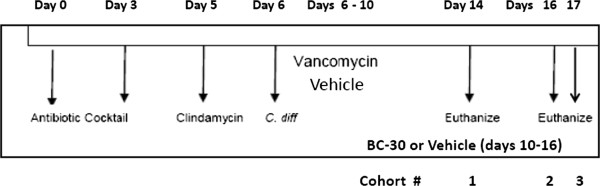
Background: Recently, we found that the probiotic strain Bacillus coagulans GBI-30, 6086 (GanedenBC30) improved indices of Clostridium difficile (C. difficile)-induced colitis in mice (Fitzpatrick et al., Gut Pathogens, 2011). Our goal was to determine if BC30 could also prevent the recurrence of C. difficile-induced colitis in mice, following initial treatment with vancomycin. During study days 0 through 5, mice were treated with antibiotics. On day 6, the C. difficile strain VPI 10463 was given by oro-gastric gavage at ≈ 5x10⁴ CFU to induce colitis. Mice were treated on study days 6 to 10 with vancomycin (50 mg/kg) (vanco) or vehicle (saline) by gavage. On days 10 to16, mice were dosed by gavage with saline vehicle or BC30 (2 x 10⁹ CFU per day). Mice were monitored for mortality, weight loss and diarrhea. On study days 14, 16 and 17, stools and colons were collected for analyzing other parameters of colitis.

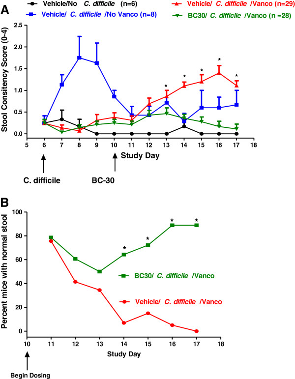
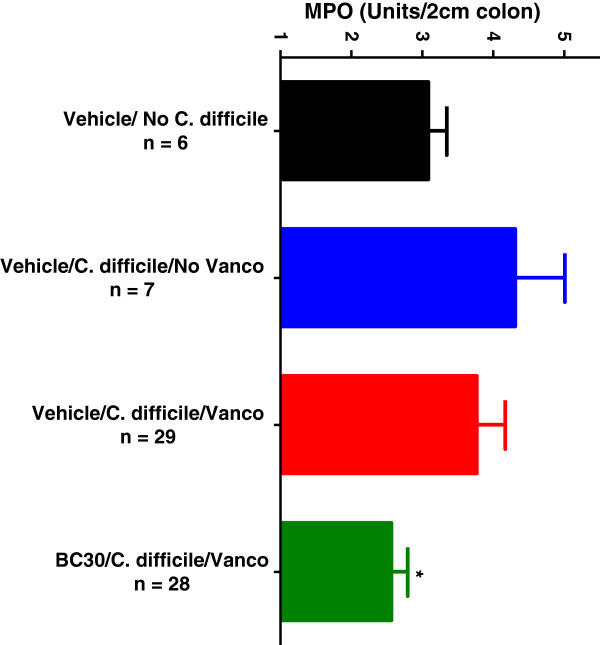
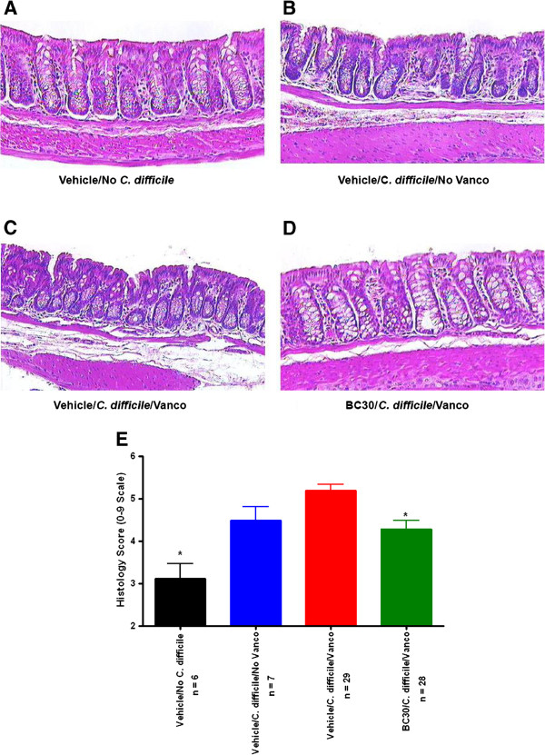
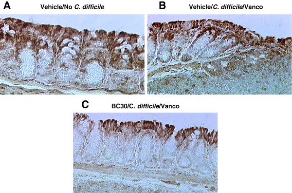
Results: The mean stool consistency score in Vehicle/C.difficile/Vanco mice increased from 0.4 (day 10) to a range of 1.1 to 1.4 (days 14 to 17), indicating the recurrence of colitis. On days 13 through 17, the stool consistency scores for the vancomycin/BC30 mice were significantly lower (p< 0.05) than for the vancomycin/vehicle cohort of animals. On day 17, 88.9% of mice treated with BC30 had normal stools, while this value was 0% with vehicle treatment (p value = 0.0004). Colonic myeloperoxidase (Units/2 cm colon) was significantly (p < 0.05) reduced from 4.3 ± 0.7 (Vehicle/C.difficile/Vanco) to 2.6 ± 0.2 (BC30/C. Difficle/Vanco). The colonic histology score and Keratinocyte derived-chemokine level in the colon were also lower in BC30 treated mice.

Summary: In BC30-treated mice, there was evidence of better stool consistency, as well as improved biochemical and histological indices of colitis, following initial treatment of animals with vancomycin.

Conclusion: BC30 limited the recurrence of CD-induced colitis following vancomycin withdrawal in mice.


Figures

Figure 1
Figure 1
Study overview. The key events associated with the Clostridium difficile induced colitis mouse model are shown. On study days 0 through 3, C57BL/6 mice received an antibiotic mixture of kanamycin, gentamicin, colistin, metronidazole and Vanco in the drinking water, followed by clindamycin (10 mg/kg, i.p., on day 5). On day 6, the C. difficile strain VPI 10463 was given by oro-gastric gavage at ≈ 5x10⁴ CFU to induce colitis. Mice were treated on study days 6 to 10 with Vanco (50 mg/kg) or vehicle (saline) by gavage. On days 10 to 16, mice were dosed by gavage with vehicle (50% maltodextrin/saline, n=29) or BC30 (2 x 10⁹ CFU per day, n=28). One negative control group of mice (n=6) was dosed with vehicle, but did not receive C. difficile, while a positive control group (initial n of 8) received C. difficile but not Vanco. Mice were monitored daily (days 6 to 17) for mortality, weight loss and stool consistency. On study days 14, 16 and 17, stools and colons were collected for further analyses.
Figure 2
Figure 2
Infection and toxin data. A) The percentages of animals positive for C. difficile in the stool were determined by ELISA on study day 14, 16 and 17. * indicates p< 0.05 vs. all other C. difficile infection groups. B) Toxin A/B levels were determined in a semi-quantitative fashion with an appropriate ELISA kit, as described in the Methods section. The values in the graph represent absorbance readings at 450 nm. * indicates p< 0.05 vs. all other C. difficile infection groups.
Figure 3
Figure 3
Stool consistency data. A) Mice were randomized on study day 6 to one of four treatment groups. All mice in the negative control group (n=6) that did not receive C. difficile (black symbols, lines) generally had normal stools throughout the study. Stool consistency scores were higher in the group of mice (blue symbols, bars) that were treated with Vehicle/C. difficile/No Vanco. In these animals, disease was prominently present on days 7 to 9. For the other two experimental groups, mice received Vehicle/C. difficile/Vanco and either Vehicle (red symbols, lines) to induce disease recurrence (study days 11 to 17), or BC30 at a dose of 2 x 10⁹ CFU per day (green symbols, lines). * indicates p < 0.05 vs. BC30/C. difficile/Vanco treatment group on study days 13 through 17. B) The percentages of mice with normal stools in the Vehicle/C. difficile/Vanco (red symbols, lines) and BC30/C. difficile/Vanco (green symbols/lines) treatment groups is shown in this panel. Data are shown for study days 10 through 17. On days 14 through 17, significant differences (* p < 0.05) were found in the percentages of mice with normal stools in the Vehicle/BC30/Vanco group as compared to the Vehicle/C. difficile/Vanco group. On day 17, 88.9% of mice treated with BC30 had normal stools while this value was 0% with vehicle treatment.
Figure 4
Figure 4
Colonic myeloperoxidase. Measurements of colonic myeloperoxidase (MPO) levels for all mice are shown as Units per 2 cm of colon. Colonic MPO was significantly (p < 0.05) reduced from 4.3 ± 0.7 (Vehicle/Vanco, red bar) to 2.6 ± 0.2 (BC30/Vanco, green bar).
Figure 5
Figure 5
Colonic histology. Representative histology pictures from hematoxylin and eosin (H&E) stained colonic specimens are shown at a magnification of 200-fold. A) A relatively normal histological appearance is evident in the colon from a mouse not infected with C. difficile.B) Evidence of crypt damage, submucosal edema and the influx of inflammatory cells in the lamina propria and sub-mucosa is present in the colon of an animal infected with C. difficile but not treated with Vanco. C) In the colon of a mouse given C. difficile plus Vanco, there is evidence of crypt disruption, leukocyte influx and prominent sub-mucosal edema. D) Mild pathology is observed in the colon of a BC30 treated mouse that was also given C. difficile plus Vanco. Modest leukocyte influx is present in the lamina propria, as well as limited sub-mucosal edema, when compared to the vehicle control (compare panels C and D). E) This panel shows a summary of the colonic histology score data. * p < 0.05 vs. Vehicle/C. difficile/Vanco treatment group (compare red and green bars in the graph).
Figure 6
Figure 6
Colonic COX-2 immunohistochemistry. COX-2 immunohistochemistry was performed on representative histology slides from colonic samples of three treatment groups (panels A, B and C). As shown in panel A, Immuno-staining for COX-2 was evident primarily in the colonic epithelial cells from a mouse that was not infected with C. difficile. In the colon of a Vehicle/C.difficile/Vanco treated animal there was prominent brown COX-2 staining in colonocytes, as well as infiltrating leukocytes within the lamina propria and submucosa (panel B). Only minimal COX-2 immuno-staining (i.e., primarily in surface colonic epithelial cells) was present within the colon of a BC30/C. difficile/Vanco treated mouse (panel C).

References

    1. McDonald LC, Kilgore GE, Thompson A, Owens RC, Kazavoka SV, Sambol SP, Johnson S, Gerding DN. An epidemic, toxin gene-variant strain of Clostridium difficile. N Engl J Med. 2005;353:2433–2441. doi: 10.1056/NEJMoa051590. - DOI - PubMed
    1. Pepin J, Valiquetet L, Alary ME, Raiche E, Ruel J, Fulop K, Godin D, Bourassa C. Clostridium-difficile-associated diarrhea in a region of Quebec from 1991–2003: a changing pattern of disease activity. CMAJ. 2004;171:466–472. doi: 10.1503/cmaj.1041104. - DOI - PMC - PubMed
    1. Johnson S, Gerding DN. Clostridium difficile-associated diarrhea. Clin Infect Dis. 1998;26:1027–1034. doi: 10.1086/520276. - DOI - PubMed
    1. Kelly CP, Pothoulakis C, Lamont JT. Clostridium difficile colitis. N Eng J Med. 1994;330:257–262. doi: 10.1056/NEJM199401273300406. - DOI - PubMed
    1. Pothoulakis C, Lamont JT. Microbes and microbial toxins: paradigms for microbial-mucosal interactions II. The integrated response of the intestine to Clostridium difficile toxins. Am J Gastrointest Liver Physiol. 2001;28:G178–G183. - PubMed

LinkOut - more resources