. 2012 Nov;4(11):927-33.
doi: 10.1038/nchem.1433. Epub 2012 Sep 9.

Amyloid β-sheet mimics that antagonize protein aggregation and reduce amyloid toxicity

Affiliations

Amyloid β-sheet mimics that antagonize protein aggregation and reduce amyloid toxicity

Pin-Nan Cheng et al. Nat Chem. 2012 Nov.

Abstract

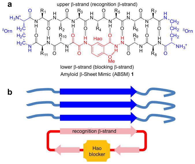
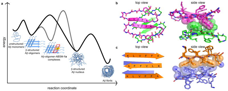
The amyloid protein aggregation associated with diseases such as Alzheimer's, Parkinson's and type II diabetes (among many others) features a bewildering variety of β-sheet-rich structures in transition from native proteins to ordered oligomers and fibres. The variation in the amino-acid sequences of the β-structures presents a challenge to developing a model system of β-sheets for the study of various amyloid aggregates. Here, we introduce a family of robust β-sheet macrocycles that can serve as a platform to display a variety of heptapeptide sequences from different amyloid proteins. We have tailored these amyloid β-sheet mimics (ABSMs) to antagonize the aggregation of various amyloid proteins, thereby reducing the toxicity of amyloid aggregates. We describe the structures and inhibitory properties of ABSMs containing amyloidogenic peptides from the amyloid-β peptide associated with Alzheimer's disease, β(2)-microglobulin associated with dialysis-related amyloidosis, α-synuclein associated with Parkinson's disease, islet amyloid polypeptide associated with type II diabetes, human and yeast prion proteins, and Tau, which forms neurofibrillary tangles.


Conflict of interest statement

The authors declare no conflict.

Figures

Figure 1
Figure 1
Design of amyloid β-sheet mimic 1. (a) Representation of ABSM 1 illustrating the upper β-strand (recognition β-strand), the δ-linked ornithine (δOrn) turn unit, and the Hao amino acid blocker unit. (b) Representation of ABSM 1 recognizing and blocking amyloid aggregation through β-sheet interactions.
Figure 2
Figure 2
X-ray crystallographic structure of ABSM 1r, which contains the heptapeptide sequence AIIGLMV (Aβ30–36). (a) The monomer. (b) The dimer: top view. (c) The dimer: side view. (d) Stacked layers of dimer in the crystal lattice. Note that the view in b is perpendicular to the β-sheet (top view), whereas the view in c and d is 90° away, parallel to the β-sheet (side view) and shows the hydrophobic contacts. Some side chains in c and d have been omitted for clarity.
Figure 3
Figure 3
Effect of ABSMs on inhibition of Aβ40, Aβ42, hβ2M, and hαSyn1–100 aggregation monitored by thioflavin T fluorescence assays and transmission electron microscopy. (a) Lag time of Aβ40 (20 μM) aggregation in the absence and presence of ABSM 1a. (b) Lag time of Aβ42 (20 μM) aggregation in the absence and presence of ABSM 1a. (c) Lag time of hβ2M (30μM) aggregation in the absence and presence of ABSM 1m. (d) Lag time of hαSyn1–100 (50μM) aggregation in the absence and presence of ABSM 1o. (e) TEM of Aβ40 (20 μM) after incubation for 6 h without ABSM 1a (top) and incubation for 6 h with 1.0 equivalent of ABSM 1a (bottom). (f) TEM of Aβ42 (20 μM) after incubation for 7 h without ABSM 1a (top) and incubation for 7 h with 1.0 equivalent of ABSM 1a (bottom). (g) TEM of hβ2M (30 μM) after incubation for 2 h without ABSM 1m (top) and incubation for 2 h with 1.0 equivalent of ABSM 1m (bottom). (h) TEM of hαSyn1–100 (50 μM) after incubation for 72 h without ABSM 1o (top) and incubation for 72 h with 1.0 equivalent of ABSM 1o (bottom). * For explanation, see the text and Figures S1, S2, and S4. Error bars correspond to standard deviation of four or greater sets of experiments. For experimental details, see the Supplementary Information Text.
Figure 4
Figure 4
Effect of ABSM 1a on Aβ40 and Aβ42 toxicity toward PC-12 cells. Addition of Aβ decreases cell survival when PC-12 cells are cultured for 24 h with preincubated Aβ. Cell survival increases when cells are cultured for 24 h with a preincubated mixture of ABSM 1a and Aβ in 0.2, 1.0, and 5 molar ratios. Cell survival is given as a percentage relative to controls in which only phosphate-buffered saline (PBS) is added. The cell survival of the PBS controls is taken to be 100%. Error bars correspond to standard deviation of four sets of experiments. For experimental details, see the Supplementary Information Text.
Figure 5
Figure 5
β-Sheet interactions of Aβ peptides and ABSM 1a. (a) Proposed model of inhibition of Aβ aggregation by ABSM 1a. The solid curve corresponds to a pathway in which Aβ aggregates without ABSM 1a. The dashed curve corresponds to an alternative pathway in which ABSM 1a inhibits Aβ aggregation by binding Aβ oligomers. (b) Crystal structure of a macrocyclic peptide containing pentapeptide sequence LVFFA. (PDB ID: 3Q9H) The magenta and green structures correspond to parallel and antiparallel β-sheet dimers formed by the macrocyclic peptide. The side view shows hydrophobic contacts formed between the parallel and antiparallel β-sheet dimers. (c) Crystal structure of the linear peptide KLVFFA. (PDB ID: 3OW9) The orange and purple structures correspond to different layers within the crystal structure. The side view shows hydrophobic contacts between the layers.

Similar articles

Cited by

References

    1. Chiti F, Dobson CM. Protein misfolding, functional amyloid, and human disease. Annu Rev Biochem. 2006;75:333–366. - PubMed
    1. Aguzzi A, O’Connor T. Protein aggregation diseases: pathogenicity and therapeutic perspectives. Nat Rev Drug Discov. 2010;9:237–248. - PubMed
    1. Bartolini M, Andrisano V. Strategies for the inhibition of protein aggregation in human diseases. ChemBioChem. 2010;11:1018–1035. - PubMed
    1. Greenwald J, Riek R. Biology of amyloid: structure, function, and regulation. Structure. 2010;18:1244–1260. - PubMed
    1. Tycko R. Solid-state NMR studies of amyloid fibril structure. Annu Rev Phys Chem. 2011;62:279–299. - PMC - PubMed

Publication types