. 2013 Apr;6(1):91-107.
doi: 10.1007/s12307-012-0121-z. Epub 2012 Oct 23.

Transcription factor networks in invasion-promoting breast carcinoma-associated fibroblasts

Affiliations

Transcription factor networks in invasion-promoting breast carcinoma-associated fibroblasts

A Siletz et al. Cancer Microenviron. 2013 Apr.

Abstract

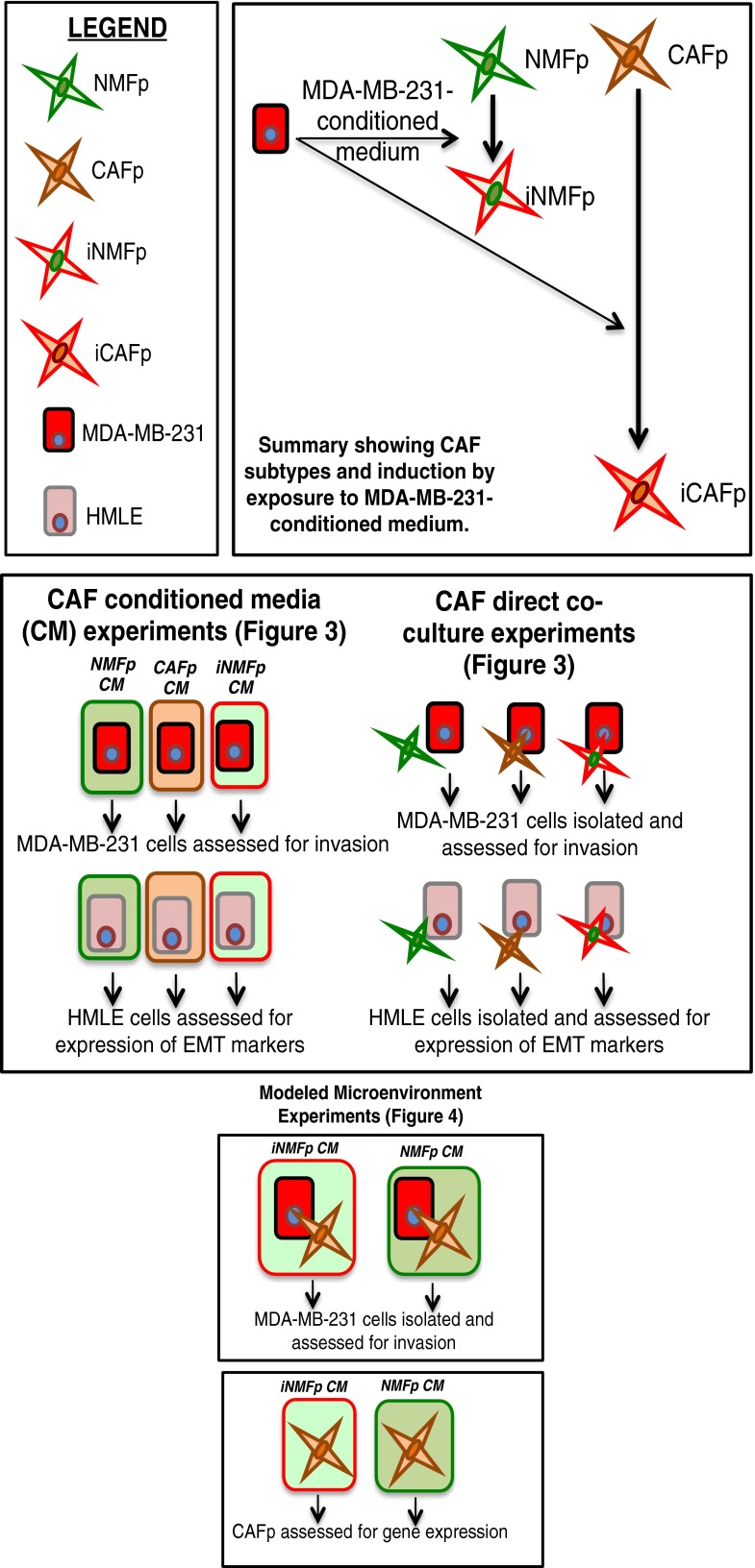
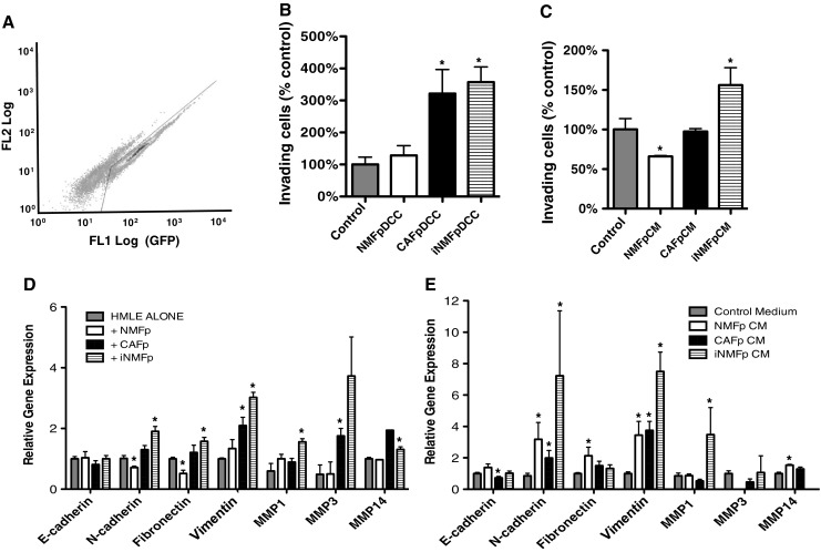
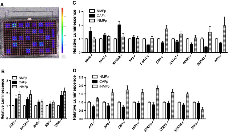
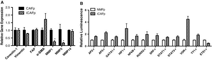
Carcinoma-associated fibroblasts (CAFs) contribute to both tumor growth and cancer progression. In this report, we applied an emerging transcription factor (TF) activity array to fibroblasts to capture the activity of the intracellular signaling network and to define a signature that distinguishes mammary CAFs from normal mammary fibroblasts. Normal fibroblasts that restrained cancer cell invasion developed into an invasion-promoting CAF phenotype through exposure to conditioned medium from MDA-MB-231 breast cancer cells. A myofibroblast-like CAF cell line expressing high levels of smooth muscle actin was compared to normal mammary fibroblasts before and after induction. Comparison of TF activity profiles for all three fibroblast types identified a TF activity signature common to CAFs which included activation of reporters for TFs ELK1, GATA1, retinoic acid receptor (RAR), serum response factor (SRF), and vitamin D receptor (VDR). Additionally, CAFs resembling myofibroblasts, relative to normal fibroblasts, had elevated activation corresponding to NF-kappaB, RUNX2, and YY1, and distinct activity patterns for several differentiation-related TF reporters. Induction of CAFs by exposure of normal fibroblasts to conditioned medium from MDA-MB-231 cells resulted in increased activation of reporters for HIF1, several STAT TFs, and proliferation-related TFs such as AP1. Myofibroblast-like CAFs and induced normal mammary fibroblasts promoted invasion of breast cancer cells by distinct mechanisms, consistent with their distinct patterns of TF activation. The TF activity profiles of CAF subtypes provide an overview of intracellular signaling associated with the induction of a pro-invasive stroma, and provide a mechanistic link between the microenvironmental stimuli and phenotypic response.


Figures

Fig. 1
Fig. 1
Schematic of experiments in this study to assess CAF subtypes and normal fibroblasts
Fig. 2
Fig. 2
Phenotypic differences between normal and carcinoma-associated fibroblasts. a Morphology of immortalized normal and carcinoma-associated mammary fibroblasts. NMF pBabe p53 (NMFp) have elongated spindle morphology and orient with long axes in the same direction. CAF pBabe p53 (CAFp) are broader, more pleiomorphic, and do not display alignment in the same direction. NMFp exposed to conditioned medium from cancer cells for 14 days to induce fibroblast activation (iNMFp) have an intermediate phenotype with broader cell bodies than NMFp and some loss of orientation of long axes. b Expression of fibroblast marker vimentin and myoepithelial marker keratin 5 in NMFp and CAFp relative to the dermal fibroblast line BJ HFF. Keratin 5 was undetectable in all fibroblast samples. c and d Gene expression of cancer-associated fibroblast markers in NMFp, CAFp, and iNMFp expressed relative to NMFp levels. Note: levels of expression of alpha smooth muscle actin in CAFp varied from 5-fold to 200-fold in all samples assayed. Values shown are representative cultures with intermediate expression. Note log scale in d. Scale bar: 100 μm. Error bars indicate standard error of the mean. Asterisk indicates significantly different from NMFp (p ≤ 0.05). n ≥ 3 for all experiments
Fig. 3
Fig. 3
Quantification of CAF secreted factors. Levels of stromal cell-derived factor 1α (SDF1α) (a) and transforming growth factor β1 (TGF-β1) (b) in medium from fibroblasts measured by enzyme-linked immunosorbent assay after subtraction of background level from fetal bovine serum in growth medium. c Levels of prostaglandin E2 (PGE2) in medium from fibroblasts measured by competitive immunoassay after subtraction of background level from fetal bovine serum in growth medium and non-specific binding effect. d Quantification of collagen I/III accumulation in fibroblast cultures measured by retention of picrosirius red dye. Staining intensity is shown as a percentage of NMFp average intensity. Representative picrosirius red staining in culture wells is shown in e. Asterisk indicates significantly different from NMFp with p ≤ 0.05. n ≥ 3 for all experiments
Fig. 4
Fig. 4
CAFp and iNMFp fibroblasts increase invasion of mammary epithelial cells by different mechanisms while NMFp fibroblasts restrain invasion by a paracrine mechanism. MECs were co-cultured directly with fibroblasts and then isolated by FACS before assaying, or cultured in conditioned media for 5 days. a Representative FACS plot showing isolation of GFP+ mammary epithelial cells from fibroblast populations. Image shown is CAFp/MDA-MB-231 co-culture with 70 % GFP+ MDA-MB-231 cells; because of the higher growth rate of mammary epithelial cells relative to fibroblasts, by the end of culture typically 70–90 % of cells in fibroblast/mammary epithelial cell co-cultures were GFP+ epithelial cells. Note typical high autofluorescence of fibroblast population (left gate) (FL1 is non-GFP fluorescence). For direct co-culture (b and d), controls were MECs cultured alone. b Relative invasion of MDA-MB-231 adenocarcinoma cells in direct co-culture (abbreviated DCC) with fibroblasts after isolation from fibroblasts using FACS. c Relative invasion of MDA-MB-231 adenocarcinoma cells cultured in conditioned media (abbreviated CM) from fibroblasts. d Expression of epithelial and mesenchymal markers by quantitative PCR in immortalized mammary epithelial (HMLE) cells after direct co-culture with fibroblasts. e Expression of epithelial and mesenchymal markers by quantitative PCR in HMLE cells exposed to conditioned media from fibroblasts. Error bars indicate standard error of the mean. Asterisk indicates significantly different relative to control with p ≤ 0.05. n ≥ 3 for all experiments
Fig. 5
Fig. 5
Combined effects of distinct CAF subtypes in a model of the tumor microenvironment. a Effect on invasiveness of MDA-MB-231 cells in direct co-culture with CAFp fibroblasts with and without addition of conditioned medium from iNMFp fibroblasts. Predicted additive effect is the sum of effects of CAFp direct co-culture alone and conditioned medium from iNMFp alone. b Changes in α-SMA mRNA expression in CAFp fibroblasts upon treatment with conditioned media from either NMFp or iNMFp fibroblasts. c Relative picrosirius red staining intensity indicative of collagen accumulation in CAFp fibroblast cultures treated with conditioned media from NMFp or iNMFp fibroblasts. Error bars indicate standard error of the mean. Asterisk indicates significantly different from control (CAFp cultured in unconditioned medium) with p ≤ 0.05. n ≥ 3 for all experiments
Fig. 6
Fig. 6
Transcription factor activity differences in invasion-promoting fibroblast subtypes versus invasion-restraining normal mammary fibroblasts. a False color image of a TF reporter array showing luminescent signal from cells transduced with TF reporters with addition of D-luciferin substrate. b Transcription factor reporters displaying significantly (p ≤ 0.05) altered activity relative to normal mammary fibroblasts in both CAFp and iNMFp. c Transcription factor reporters displaying significantly (p ≤ 0.05) altered activity relative to normal mammary fibroblasts in CAFp not observed in iNMFp. Reporters with increased activity in CAFp are shown in alphabetical order followed by reporters with decreased activity in alphabetical order. d Transcription factor reporters displaying significantly (p ≤ 0.05) altered activity relative to normal mammary fibroblasts in iNMfp not observed in CAFp. Normalized luminescence proportional to transcription factor-dependent translation of firefly luciferase is shown on y-axes. Note E2F1-r activity was significantly different from NMFp in both CAFp and iNMFp but changes were in opposite directions as shown in c and d. n ≥ 3 for all experiments
Fig. 7
Fig. 7
Gene expression changes and TF activity in CAFp fibroblasts exposed to cancer cell-conditioned medium. a Quantitative PCR for CAFp fibroblasts treated with medium conditioned by MDA-MB-231 breast adenocarcinoma cells (iCAFp) compared to untreated CAFp. Compare with Fig. 2c and d. Error bars indicate standard error of the mean. Asterisk indicates p ≤ 0.05. n = 3. b Transcription factor reporters displaying significantly (p ≤ 0.05) altered activity relative to normal mammary fibroblasts in iCAFp. Normalized luminescence proportional to transcription factor-dependent translation of firefly luciferase is shown on y-axis. n = 3
Fig. 8
Fig. 8
Venn diagram summarizing TF reporter activation compared to NMFp in CAF subtypes. *E2F1-r activity was significantly elevated in iNMFp compared to NMFp fibroblasts. The increased activity seen in iCAFp was not significantly different from NMFp levels, but was significantly greater than parental CAFp levels. Significant elevation of E2F1-r activity relative to baseline is therefore a common response of iNMFp and iCAFp fibroblasts to MDA-MB-231 conditioned medium, and the opposite is seen in CAFp. Note ELK1-r and RAR-r activities were nonsignificantly elevated relative to NMFp in iCAFp fibroblasts; for ELK1-r p = 0.06 and for RAR-r p = 0.08. For all other TF reporters shown p ≤ 0.05 for all comparisons

Similar articles

Cited by

References

    1. Hanahan D, Weinberg RA. Hallmarks of cancer: the next generation. Cell. 2011;144(5):646–674. doi: 10.1016/j.cell.2011.02.013. - DOI - PubMed
    1. Allinen M, Beroukhim R, Cai L, Brennan C, Lahti-Domenici J, Huang H, Porter D, Hu M, Chin L, Richardson A, Schnitt S, Sellers WR, Polyak K. Molecular characterization of the tumor microenvironment in breast cancer. Cancer Cell. 2004;6(1):17–32. doi: 10.1016/j.ccr.2004.06.010. - DOI - PubMed
    1. Barcellos-Hoff MH, Ravani SA. Irradiated mammary gland stroma promotes the expression of tumorigenic potential by unirradiated epithelial cells. Cancer Res. 2000;60(5):1254–1260. - PubMed
    1. Bissell MJ, Radisky D. Putting tumours in context. Nat Rev Cancer. 2001;1(1):46–54. doi: 10.1038/35094059. - DOI - PMC - PubMed
    1. Kalluri R, Zeisberg M. Fibroblasts in cancer. Nat Rev Cancer. 2006;6(5):392–401. doi: 10.1038/nrc1877. - DOI - PubMed

LinkOut - more resources