. 2013 Feb;24(2):343-54.
doi: 10.1007/s10856-012-4793-1. Epub 2012 Oct 23.

Nanoparticles of cobalt-substituted hydroxyapatite in regeneration of mandibular osteoporotic bones

Affiliations

Nanoparticles of cobalt-substituted hydroxyapatite in regeneration of mandibular osteoporotic bones

Nenad Ignjatović et al. J Mater Sci Mater Med. 2013 Feb.

Abstract

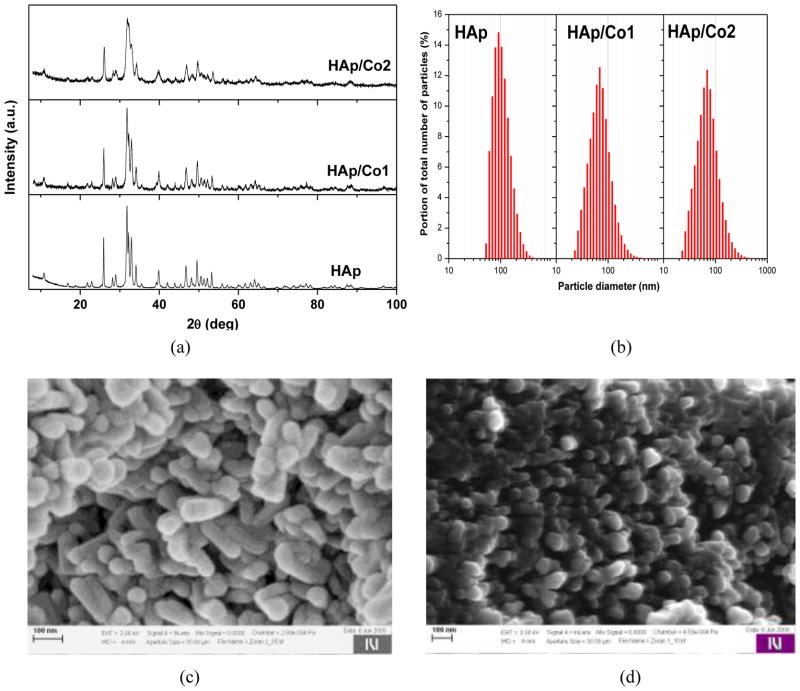
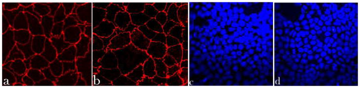
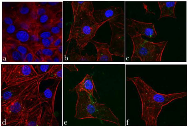
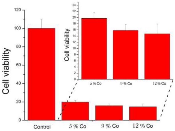
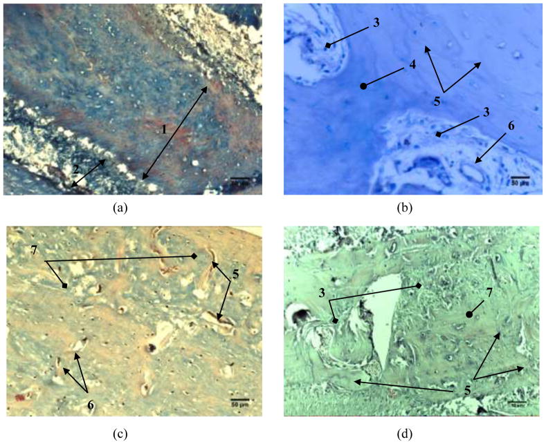
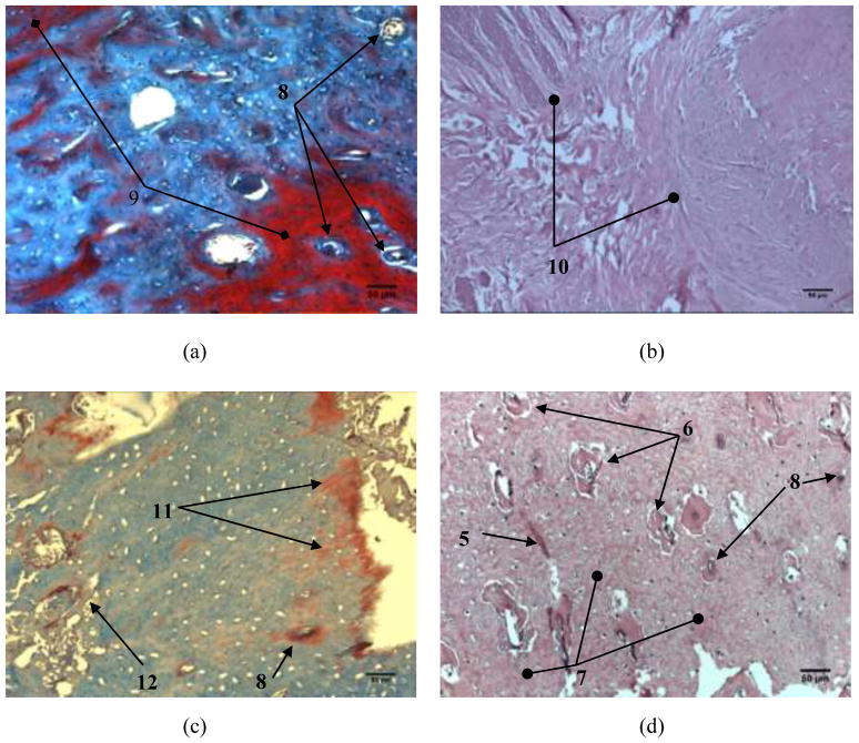
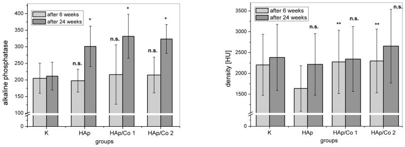
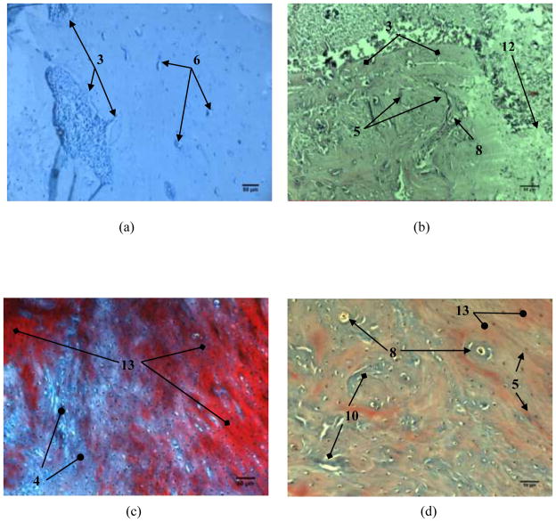
Indications exist that paramagnetic calcium phosphates may be able to promote regeneration of bone faster than their regular, diamagnetic counterparts. In this study, analyzed was the influence of paramagnetic cobalt-substituted hydroxyapatite nanoparticles on osteoporotic alveolar bone regeneration in rats. Simultaneously, biocompatibility of the material was tested in vitro, on osteoblastic MC3T3-E1 and epithelial Caco-2 cells in culture. The material was shown to be biocompatible and nontoxic when added to epithelial monolayers in vitro, while it caused a substantial decrease in the cell viability as well as deformation of the cytoskeleton and cell morphology when incubated with the osteoblastic cells. In the course of 6 months after the implantation of the material containing different amounts of cobalt, ranging from 5 to 12 wt%, in the osteoporotic alveolar bone of the lower jaw, the following parameters were investigated: histopathological parameters, alkaline phosphatase and alveolar bone density. The best result in terms of osteoporotic bone tissue regeneration was observed for hydroxyapatite nanoparticles with the largest content of cobalt ions. The histological analysis showed a high level of reparatory ability of the nanoparticulate material implanted in the bone defect, paralleled by a corresponding increase in the alveolar bone density. The combined effect of growth factors from autologous plasma admixed to cobalt-substituted hydroxyapatite was furthermore shown to have a crucial effect on the augmented osteoporotic bone regeneration upon the implantation of the biomaterial investigated in this study.


Figures

Fig. 1
Fig. 1
a) XRD patterns of synthesized materials; b) size distribution of particles obtained by LD; c) and d) SEM images of nanopowder samples HAp and HAp/Co1, respectively.
Fig. 2
Fig. 2
Immunofluorescent staining of ZO-1 molecules (a–b) and cell nucleus (c–d) in Caco-2 cell monolayers treated with either no particles (control, a, c) or 2 mg/cm2 of HAp/Co2 particles (b, d), imaged under identical excitations. The sizes of images (a, b) and (c, d) is 200 × 200 μm and 475 × 475 μm, respectively.
Fig. 3
Fig. 3
Confocal optical micrographs of fluorescently stained osteoblastic cell nucleus (blue) and cytoskeletal f-actin (red), and HAp/Co particles and/or intracellular mineral particles (green) following 7 days of incubation with either no particles (control, a) or 2 mg/cm2 of HAp/Co particles (b–f). The size of each image is 750 × 750 μm.
Fig. 4
Fig. 4
Mitochondrial activity indicative of cell viability, normalized to the negative control (C−) and determined by the MTT assay for HAp/Co particles containing different weight percentage of Co2+ ions: 5 wt% (HAp/Co1), 9 wt% and 12 wt% (HAp/Co2).
Fig. 5
Fig. 5
Histopathology of alveolar bone 6 weeks after the implantation: a) control group, b) HAp; c) HAp/Co1 and d) HAp/Co2. [labels: 1 - compact bone, 2 - cancellous bone, 3 - replacement of the material with new bone tissue, 4 - a young bone, 5 - cement lines, 6 - blood vessel, 7 - the newly formed bone]
Fig. 6
Fig. 6
Histopathology of alveolar bone 24 weeks: a) control group, b) HAp; c) HAp/Co1; d) HAp/Co2. [labels: 5 - cement lines, 6 - blood vessel, 7 - the newly formed bone, 8 - Haversian canals, 9 - bone mineralization, 10 - osteogenesis, 11 - the beginning of ossification and calcification, 12 - collagen fibers, 13 - mature calcified bone]
Fig. 7
Fig. 7
(a) ALP content (U/L) and (b) densities of alveolar bone expressed in Haunsfiled units (HU), 6 and 24 weeks after the implantation, for different experimental groups (HAp, HAp/Co1, HAp/Co2) and the control (K). K indicates healthy bone in (b). Data are shown as means with error bars representing standard deviation (* => p < 0.01 with respect to the control group; ** => p < 0.05 with respect to HAp group, n.s. => non-significant with respect to the control group).
Fig. 8
Fig. 8
The histopathology of alveolar bone reconstructed with HApCo2 after 6 (a, b) and 24 weeks (c, d). The material was mixed with (a, c) blood and (b, d) blood plasma. [labels: 3 - replacement of the material with new bone tissue, 4 - a young bone, 5 - cement lines, 6 - blood vessel, 8 - Haversian canals, 10 - osteogenesis, 12 - collagen fibers, 13 - mature calcified bone]
Fig. 9
Fig. 9
(a) ALP content (U/L) and (b) densities of alveolar bone expressed in Haunsfiled units (HU) for the control (K) and experimental groups involving the implantation of HAp/Co2 mixed with saline, blood and blood plasma, 6 and 24 weeks after the implantation. Data are shown as means with error bars representing standard deviation (* => p < 0.01 with respect to the control group; ** => p < 0.05 with respect to the control group; n.s. => non-significant with respect to the control group).

References

    1. Raisz LG. Pathogenesis of osteoporosis: concepts, conflicts, and prospects. J Clin Invest. 2005;115:3318–3325. - PMC - PubMed
    1. Pollähne W, Pfeifer M, Lazarescu A, Minne HW. Osteoporose: Bildgebende Diagnostik. Medizin im Bild. 1996;3:37–44.
    1. Marco F, Milena F, Gianluca G, Vittoria O. Peri-implant osteogenesis in health and osteoporosis. Micron. 2005;36(7–8):630–644. - PubMed
    1. Gupta AK, Gupta M. Synthesis and surface engineering of iron oxide nanoparticles for biomedical applications. Biomaterials. 2005;26(18):3995–4021. - PubMed
    1. Ito A, Honda H, Kobayasji T. Cancer immunotherapy based on intracellular hyperthermia using magnetite nanoparticles: a novel concept of “heat-controlled necrosis” with heat shock protein expression. Cancer Immunol Immunother. 2006;55(3):320–328. - PMC - PubMed

Publication types

MeSH terms