. 2012;10(10):e1001407.
doi: 10.1371/journal.pbio.1001407. Epub 2012 Oct 16.

Generation of functional blood vessels from a single c-kit+ adult vascular endothelial stem cell

Affiliations

Generation of functional blood vessels from a single c-kit+ adult vascular endothelial stem cell

Shentong Fang et al. PLoS Biol. 2012.

Abstract

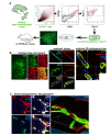
In adults, the growth of blood vessels, a process known as angiogenesis, is essential for organ growth and repair. In many disorders including cancer, angiogenesis becomes excessive. The cellular origin of new vascular endothelial cells (ECs) during blood vessel growth in angiogenic situations has remained unknown. Here, we provide evidence for adult vascular endothelial stem cells (VESCs) that reside in the blood vessel wall endothelium. VESCs constitute a small subpopulation within CD117+ (c-kit+) ECs capable of undergoing clonal expansion while other ECs have a very limited proliferative capacity. Isolated VESCs can produce tens of millions of endothelial daughter cells in vitro. A single transplanted c-kit-expressing VESC by the phenotype lin-CD31+CD105+Sca1+CD117+ can generate in vivo functional blood vessels that connect to host circulation. VESCs also have long-term self-renewal capacity, a defining functional property of adult stem cells. To provide functional verification on the role of c-kit in VESCs, we show that a genetic deficit in endothelial c-kit expression markedly decreases total colony-forming VESCs. In vivo, c-kit expression deficit resulted in impaired EC proliferation and angiogenesis and retardation of tumor growth. Isolated VESCs could be used in cell-based therapies for cardiovascular repair to restore tissue vascularization after ischemic events. VESCs also provide a novel cellular target to block pathological angiogenesis and cancer growth.


Conflict of interest statement

The authors have declared that no competing interests exist.

Figures

Figure 1
Figure 1. Isolated adult lin−CD31+CD105+ ECs encompass rare endothelial CFCs.
(A) Quantification of colony-forming ability of lin−CD31+CD105+ cells isolated from the mouse lungs. Freshly isolated cells from ten mice were assayed in duplicate. An EC colony in the semi-solid matrix is also shown. The colonies grow beneath the methylcellulose matrix adhered to the bottom of the culture dish. Scale bar, 150 µm. (B) Freshly isolated GFP-tagged ECs were seeded on standard 2-D EC cultures one CFU per culture dish together with 20 CFUs of wt ECs and grown until the monolayer was confluent. GFP+ cells form a single circular EC batch within the confluent wt EC monolayer, demonstrating the clonal growth pattern of the ECs responsible for creating the confluent monolayer. DAPI and bright field images show also the wt ECs in the confluent monolayer. Scale bar, 200 µm. (C) Colonies formed from isolated lin−CD31+CD105+ mouse lung ECs in low-cell density adherent semi-solid methylcellulose matrix were stained for various cell-surface markers. The colonies express EC markers CD31, CD105, VE-cadherin, and vWF while the cells are negative for the pan-hematopoietic marker CD45. The nuclei are stained with DAPI (blue) to recognize individual cells. MS-1 murine EC line was used as a control. Rabbit anti-β-gal antibodies and rat anti-mouse CD45 antibodies provide the isotype controls for rabbit and rat antibodies, respectively. Scale bars, 50 µm.
Figure 2
Figure 2. Isolated colony-forming ECs produce tens of millions of endothelial daughter cells in vitro.
(A) Growth kinetics of five separate EC monolayer cultures originating from individual lin−CD31+CD105+ CFCs that were picked up from colony assays and propagated in 2-D cultures. The cultures were split when 90% confluent and no cells were discarded during the experiment. A summary growth curve (mean ± SD) of the five cultures is also shown. Cell number is in log scale. (B) The long term 2-D EC cultures were analyzed at passage 24 by immunofluorescence microscopy. The cells express the endothelial markers CD105, VEGFR-2, and the stem/progenitor cell marker CD117. The nuclei are stained with DAPI (blue) to recognize individual cells. Scale bars, 50 µm; 5 µm (insert).
Figure 3
Figure 3. The CD117+ EC population enriches for rare adult colony-forming ECs.
(A) The relative distributions and overlap of CD105+, Sca-1+, and CD117+ subpopulations within isolated mouse lung lin−CD31+ ECs are shown. The results (mean ± SD) are from six FACS analyses of six mice. (B) Comparison of CFCs within the CD117-depleted and CD117-enriched fractions of lin−CD31+CD105+ ECs in vitro in colony-forming assays. Practically all CFCs are encompassed within the CD117+ EC population (p<0.0001, the Mann-Whitney test). The horizontal lines indicate 10th, 25th, 50th (median), 75th, and 90th percentiles. The results of four independent experiments, each performed in duplicate, are shown. (C) A total of 960 freshly isolated GFP+ lin−CD31+CD105+Sca1+CD117+ single cells were sorted into individual wells of 96-well plates together with a carrier population of 2,500 wt (GFP−) lin−CD31+CD105+ cells per well. At day 7, most (99.4%) of the generated EC monolayers contained only a few GFP+ ECs (above). On six wells (0.6%), the generated monolayer contained a circular, clonal area of GFP+ ECs within the otherwise GFP− monolayer (below). The result thus again reveals a small subpopulation within CD117+ ECs that are capable of undergoing clonal expansion while the other ECs have a very limited proliferative capacity. Scale bar, 200 µm. (D) Freshly isolated murine lung lin−CD31+CD105+Sca1+CD117+ ECs were assayed for a large set of endothelial, hematopoietic, and smooth muscle cell-surface markers using FACS. The results show that isolated lin−CD31+CD105+Sca1+CD117+ are highly immunoreactive for various endothelial markers while no immunoreactivity is detected against specific hematopoietic markers or against smooth muscle α-actin (SMA). The numbers indicate the mean fluorescence intensity (MFI) values (±SD) of the gated populations calculated from three mice over three experiments, and the corresponding p-values tested for significance in comparison to the respective IgG control. p-Values<0.05 were considered significant (Student's t test).
Figure 4
Figure 4. Quiescent vascular endothelium as well as growing neoangiogenic and tumor vessels contain a subpopulation of CD117+ ECs.
(A) CD117+ cells localize in the vascular endothelium. High resolution confocal scans from CD31/CD117/DAPI co-staining of mouse lung capillaries are shown. Bright field CD117 immunohistochemistry stainings are also shown. Note the red blood cells at the vessel lumina that is located adjacent to the CD117+ ECs. Scale bars, 10 µm. (B) Neoangiogenic vessels within subcutaneous matrigel plugs and B16 melanoma tumors contain numerous or CD117+ (white) ECs. ECs are stained for CD31 or vWF (both red). High resolution confocal scans are shown. Scale bars, 20 µm. (C) CD117+ ECs are detected also in the tumor vasculature in human malignant melanomas and in human breast cancer. Scale bars, 25 µm. (D) The possibility that contaminating hematopoietic stem or progenitor cells are the origin of the EC colonies was studied in control experiments. No colonies were formed by isolated BM lin− cells when studied in endothelial colony-forming assays (six independent experiments performed in duplicate). BM lin− cells were separated by standard immunomagnetic lineage depletion. In standardized hematopoietic colony-forming assays BM lin− cells produced classical hematopoietic colonies, confirming their viability. Lung lin−CD31+CD105+Sca1+CD117+ ECs formed only EC colonies in both assay formats. Scale bars, 200 µm. (E) Two different genetic reporter systems for the gene expression of VEGFR-2 and Tie-2, receptor tyrosine kinases that are expressed by vascular ECs were used to further test the endothelial origin of the isolated CFCs. lin−CD31+CD105+Sca1+CD117+ cells were isolated from the transgenic mice and then analyzed for the activity of the lacZ-β-gal reporter system (in red fluorescence) in the formed endothelial colonies. Fluorescence and bright field channel images of the colonies are shown. Colonies from the VEGFR-2 promoter lacZ mice and from the Tie-2 lacZ mice express the reporter gene (red fluorescence). Scale bars, 200 µm.
Figure 5
Figure 5. A single c-kit-expressing adult VESC by the phenotype lin−CD31+CD105+Sca-1+CD117+ can generate functional, perfused blood vessels in vivo.
(A) Flow diagram of the FACS sorting procedure used to obtain lin−CD31+CD105+Sca1+ CD117+ cells is shown. A single clonal colony originating from a single GFP-tagged lin−CD31+CD105+Sca1+CD117+ CFC was expanded for 12 d in adherent culture to amplify the cell number, manually picked up using a micropipette, mixed with 200 µl of matrigel supplemented with VEGF and bFGF, and injected subcutaneously into wt C57BL/6J mice. An eGFP channel inverted microscope micrograph of a single colony prior that was picked up and transplanted to a wt host is also shown. Scale bar, 150 µm. (B) Functional, perfused GFP+ blood vessels generated by the transplanted descendants of a single c-kit-expressing colony-forming EC by the phenotype lin− CD31+CD105+Sca1+CD117+ cell (14 d after transplantation). The mouse was perfused with fluorescent 0.2 µm microspheres (red) to stain endothelia in functional blood vessels that are connected to the blood circulation. ECs were stained for CD31 or CD105. Scale bars, 100 µm. 1-µm thick confocal optical slices and a 3-D orthogonal projection (x–z and y–z axes) are also shown. Note the blood vessel lumina (*) and the red endothelial signal from the microsphere perfusion of functional vasculature. Scale bars, 10 µm. Six independent experiments with similar results were performed. (C) Self-renewal capacity, a defining characteristic of stem cells, was evaluated by inoculating mice with syngeneic B16 melanomas (2 million cells per mice) together with 15 CFUs of GFP-tagged isolated CD31+CD105+ ECs. After 2 wk of tumor growth, repeated isolations and serial transplantations of lineage depleted single cell suspensions containing the GFP+ tagged ECs and the B16 cells were performed. The figure shows GFP+ blood vessels in the quaternary transplant. ECs were stained for VEGFR-2 (red), vWF (white), and CD31 and CD105 (red). Scale bar, 10 µm. A 3-D reconstitution of a GFP+ blood vessel in the quaternary transplant is also shown (right; a 34-µm thick stack of 34 x–y slices from a confocal scan). Six independent experiments with similar results were performed.
Figure 6
Figure 6. The CD117+ EC population greatly enriches for ECs capable of generating functional blood vessels in vivo.
(A) CD117-depleted GFP-tagged lin−CD31+CD105+Sca-1+ ECs were transplanted in matrigel plugs (here 10,000 ECs per plug) into wt C57BL/6 mice. 14 d later, none of the plugs (n = 20) contained GFP+ blood vessels. Note that occasional GFP+ ECs from the CD117-depleted GFP+ transplant can be seen within the plug, but they are infrequent (corresponding to the transplanted cell density of 50 ECs per 1 µl) and do not form complete blood vessels. A confocal scan of an area with occasional GFP+ cells is also shown. (B) An equal number (here 10,000) of CD117-enriched GFP+ lin−CD31+CD105+Sca-1+ ECs formed GFP+ blood vessels in all the matrigel plugs (n = 12) in an identical control experiment. Overview fluorescence micrographs of the plugs and confocal scans of the sectioned pugs are shown for both groups. Scale bars, 50 µm. Note that in addition to donor GFP+ ECs from the EC transplant the matrigel plugs also contain various host-derived non-EC types such as perivascular pericytes, other mesenchymal/stromal cells, and numerous infiltrating inflammatory cells. Therefore, many cells within a plug do not express endothelial cell markers such as CD31 or CD105. Additionally, the plugs also contain numerous GFP-negative wt ECs and blood vessels from the wt host (arrow).
Figure 7
Figure 7. A genetic deficit in endothelial c-kit expression decreases colony-forming VESCs and results in impaired EC proliferation and angiogenesis, and retardation of tumor growth in vivo.
(A) Equivalent lin−CD31+CD105+ EC populations were detected in mutant C57BL/6J mice with a genetic c-kit expression deficit (C57BL/6J-KitW-sh mice) and in the wt C57BL/6J controls. However, the KitW-sh mutant mice have very low numbers of CD117+ ECs (here 1% of total lin−CD31+CD105+ ECs). Typical results from FACS analysis of lung ECs are shown. Histograms indicate the percentage of CD117+ cells, control IgG labeling and gatings are also shown. (B) lin−CD31+CD105+ ECs from kit deficient KitW-sh mutant mice contain abnormally low levels of endothelial CFCs in comparison to wt mice (p<0.0001, the Mann-Whitney test). The horizontal lines indicate 10th, 25th, 50th (median), 75th, and 90th percentiles. The results of 12 independent experiments, each performed in duplicate, are shown. The Mann-Whitney test was used to compare the groups. (C) When syngeneic B16 melanoma tumors were implanted to kit deficient KitW-sh mutant mice, a highly significant impairment of tumor angiogenesis was observed (p = 0.0006; n = 7 for each group). vWF/DAPI stains are also shown. The tumor vasculature in kit deficient KitW-sh mutant mice contained a significantly diminished number of proliferating ECs (p = 0.01; n = 7 for each group). The percentiles of mean percentages of proliferating (ki-67+) ECs are shown. ki-67/CD31/DAPI stains are also shown. The Mann-Whitney test was used to compare the groups. Scale bars, 100 µm. (D) A highly significant retardation of tumor growth was observed in the kit deficient KitW-sh mice. *p<0.01; **p<0.001; ***p<0.0001; n = 17 for each group.
Figure 8
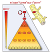
Figure 8. The present results provide evidence for adult endothelial stem cell hierarchy.
The results provide evidence for the existence of a rare self-renewing adult VESC that resides at the blood vessel wall endothelium. VESCs are a small subpopulation within vessel wall CD117+ ECs capable of undergoing clonal expansion while other ECs have a very limited proliferative capacity.

Comment in

References

    1. Coultas L, Chawengsaksophak K, Rossant J (2005) Endothelial cells and VEGF in vascular development. Nature 438: 937–945. - PubMed
    1. Eichmann A, Yuan L, Moyon D, Lenoble F, Pardanaud L, et al. (2005) Vascular development: from precursor cells to branched arterial and venous networks. Int J Dev Biol 49: 259–267. - PubMed
    1. Salven P, Mustjoki S, Alitalo R, Alitalo K, Rafii S (2003) VEGFR-3 and CD133 identify a population of CD34+ lymphatic/vascular endothelial precursor cells. Blood 101: 168–172. - PubMed
    1. Hanahan D, Weinberg RA (2011) Hallmarks of cancer: the next generation. Cell 144: 646–674. - PubMed
    1. Carmeliet P (2005) Angiogenesis in life, disease and medicine. Nature 438: 932–936. - PubMed

Publication types

Substances