

Dental approach to craniofacial syndromes: how can developmental fields show us a new way to understand pathogenesis?
- PMID: 23091490
- PMCID: PMC3467949
- DOI: 10.1155/2012/145749
Dental approach to craniofacial syndromes: how can developmental fields show us a new way to understand pathogenesis?
Abstract
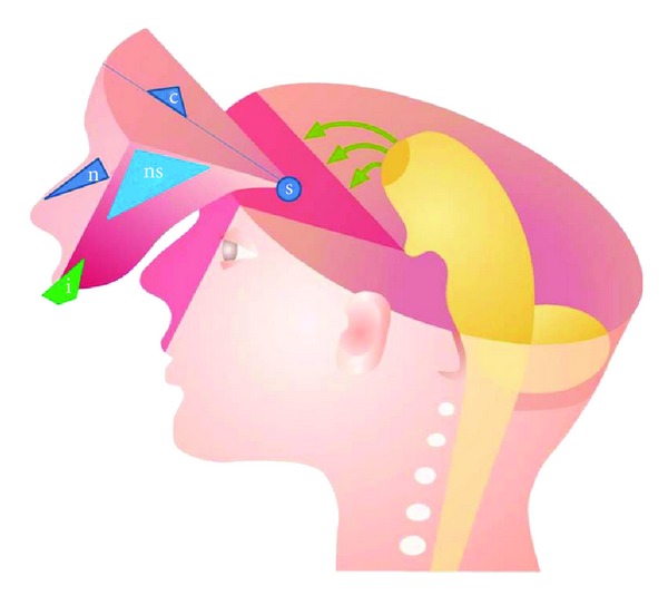
The paper consists of three parts. Part 1: Definition of Syndromes. Focus is given to craniofacial syndromes in which abnormal traits in the dentition are associated symptoms. In the last decade, research has concentrated on phenotype, genotype, growth, development, function, and treatment. Part 2: Syndromes before Birth. How can the initial malformation sites in these syndromes be studied and what can we learn from it? In this section, deviations observed in syndromes prenatally will be highlighted and compared to the normal human embryological craniofacial development. Specific focus will be given to developmental fields studied on animal tissue and transferred to human cranial development. Part 3: Developmental Fields Affected in Two Craniofacial Syndromes. Analysis of primary and permanent dentitions can determine whether a syndrome affects a single craniofacial field or several fields. This distinction is essential for insight into craniofacial syndromes. The dentition, thus, becomes central in diagnostics and evaluation of the pathogenesis. Developmental fields can explore and advance the concept of dental approaches to craniofacial syndromes. Discussion. As deviations in teeth persist and do not reorganize during growth and development, the dentition is considered useful for distinguishing between syndrome pathogenesis manifested in a single developmental field and in several fields.
Figures
Similar articles
-
Variability of the cranial and dental phenotype in Williams syndrome.Swed Dent J Suppl. 2005;(170):3-67. Swed Dent J Suppl. 2005. PMID: 15762376
-
Sella turcica morphology and the pituitary gland-a new contribution to craniofacial diagnostics based on histology and neuroradiology.Eur J Orthod. 2015 Feb;37(1):28-36. doi: 10.1093/ejo/cjs091. Epub 2012 Nov 16. Eur J Orthod. 2015. PMID: 23159420 Review.
-
Craniofacial malformations and their association with brain development: the importance of a multidisciplinary approach for treatment.Odontology. 2020 Jan;108(1):1-15. doi: 10.1007/s10266-019-00433-7. Epub 2019 Jun 6. Odontology. 2020. PMID: 31172336 Review.
-
Localised scleroderma en coup de sabre affecting the skin, dentition and bone tissue within craniofacial neural crest fields. Clinical and radiographic study of six patients.Eur Arch Paediatr Dent. 2019 Aug;20(4):339-350. doi: 10.1007/s40368-019-00427-7. Epub 2019 Mar 7. Eur Arch Paediatr Dent. 2019. PMID: 30847683
-
Update on 13 Syndromes Affecting Craniofacial and Dental Structures.Front Physiol. 2017 Dec 14;8:1038. doi: 10.3389/fphys.2017.01038. eCollection 2017. Front Physiol. 2017. PMID: 29311971 Free PMC article. Review.
Cited by
-
Binder's phenotype with ankyloglossia: Report of a rare inherited association in an Indian female.J Oral Maxillofac Pathol. 2022 Feb;26(Suppl 1):S5-S11. doi: 10.4103/jomfp.jomfp_143_21. Epub 2022 Feb 28. J Oral Maxillofac Pathol. 2022. PMID: 35450230 Free PMC article.
-
Evaluation of Twenty Non-Metric Dental Crown Traits in Different Types of Malocclusions in a Sample from India, New Delhi Population.Acta Stomatol Croat. 2023 Dec;57(4):364-380. doi: 10.15644/asc57/4/7. Acta Stomatol Croat. 2023. PMID: 38283315 Free PMC article.
-
Region-specific gene expression profiling of early mouse mandible uncovered SATB2 as a key molecule for teeth patterning.Sci Rep. 2024 Aug 6;14(1):18212. doi: 10.1038/s41598-024-68016-3. Sci Rep. 2024. PMID: 39107332 Free PMC article.
-
Mechanism of human tooth eruption: review article including a new theory for future studies on the eruption process.Scientifica (Cairo). 2014;2014:341905. doi: 10.1155/2014/341905. Epub 2014 Feb 12. Scientifica (Cairo). 2014. PMID: 24688798 Free PMC article. Review.
-
Prenatal exposure to antiepileptic drugs and dental agenesis.PLoS One. 2014 Jan 8;9(1):e84420. doi: 10.1371/journal.pone.0084420. eCollection 2014. PLoS One. 2014. PMID: 24416231 Free PMC article.
References
-
- Spranger J, Benirschke K, Hall JG. Errors of morphogenesis: concepts and terms. Recommendations of an international working group. Journal of Pediatrics. 1982;100(1):160–165. - PubMed
-
- Dorland. Dorland's Illustrated Medical Dictionary. 30th edition. Philadelphia, Pa, USA: Saunders; 2003.
-
- Gorlin RJ, Cohen MM, Jr., Levin LS. Syndrome of the Head and Neck. New York, NY, USA: Oxford University Press; 1990. (Oxford Monographs on Medical Genetics No. 19).
-
- Schalk-van der Weide Y. Oligodontia: a clinical, radiographic and genetic evaluation [thesis] Rijksuniversitet Utrecht; 1992.
-
- Kjær I. Orthodontics and foetal pathology: a personal view on craniofacial patterning. European Journal of Orthodontics. 2010;32(2):140–147. - PubMed
LinkOut - more resources
Full Text Sources
Research Materials
