

Cardiodynamics and infarct size in regional and global ischemic isolated heart model: comparison of 1 hour and 2 hours reperfusion
- PMID: 23091504
- PMCID: PMC3467443
- DOI: 10.4070/kcj.2012.42.9.600
Cardiodynamics and infarct size in regional and global ischemic isolated heart model: comparison of 1 hour and 2 hours reperfusion
Abstract
Background and objectives: We investigated whether 1 hour reperfusion is enough to assess cardiodynamics and infarct size in both regional ischemia (RI) and global ischemia (GI) in isolated rat heart models.
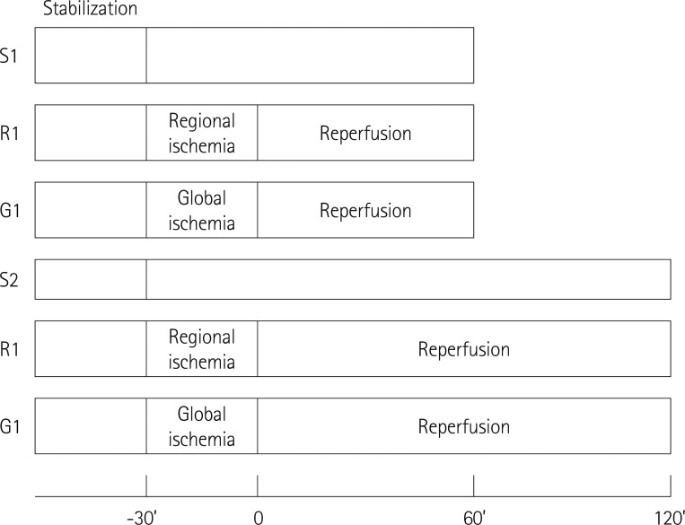
Materials and methods: Hearts were randomly assigned to one of the following groups (each n=14): 1) Sham hearts for 1 hour; 2) Sham hearts for 2 hours; 3) 30 minutes RI followed by 1 hour reperfusion; 4) 30 minutes of RI followed by 2 hours reperfusion; 5) 30 minutes GI followed by 1 hour reperfusion; and 6) 30 minutes GI followed by 2 hours reperfusion.
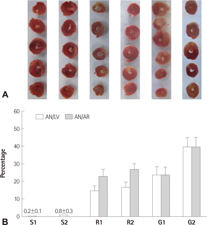
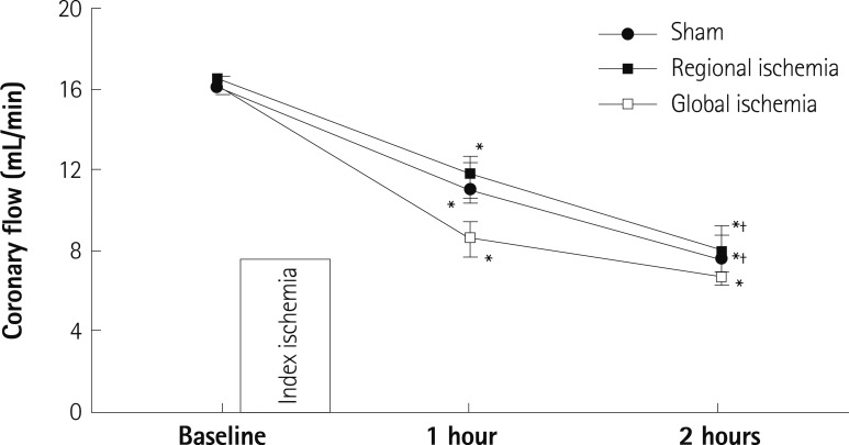
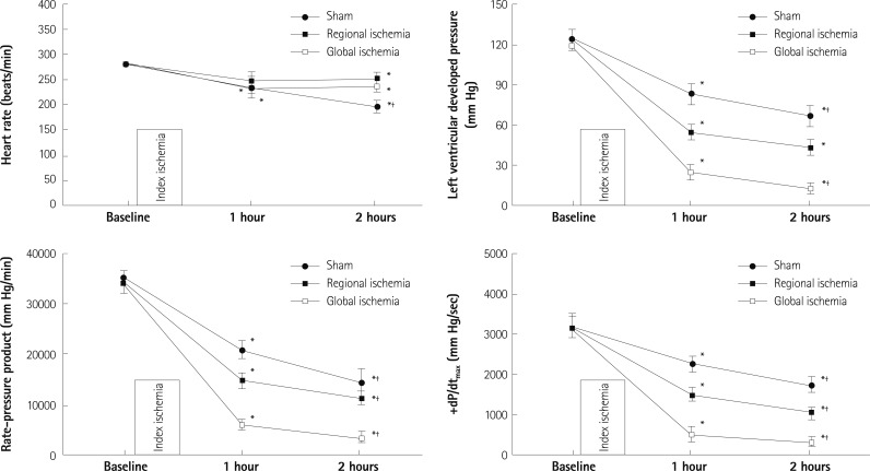
Results: There were no significant differences in infarct size between 1 hour and 2 hours reperfusion in both RI and GI. Left ventricular developed pressure was significantly decreased at both 1 hour and 2 hours reperfusion in groups of RI and GI compared to baseline (p<0.01). Rate-pressure product and +dP/dt(max) also significantly decreased compared to baseline level at both 1 hour and 2 hours reperfusion in groups of RI and GI (p<0.05).
Conclusion: There was no significant difference in infarct size between 1 hour and 2 hours reperfusion in groups of RI and GI. Cardiodynamic variables measured at 1 hour and 2 hours reperfusion significantly decreased compared to baseline level. Our data suggests that reperfusion of 1 hour is sufficient to assess cardiodynamics in both regional and global ischemic isolated hearts model.
Keywords: Heart; Myocardial infarction; Myocardial ischemia; Myocardial reperfusion.
Conflict of interest statement
The authors have no financial conflicts of interest.
Figures
Similar articles
-
Polyphenol (-)-epigallocatechin gallate targeting myocardial reperfusion limits infarct size and improves cardiac function.Korean J Anesthesiol. 2010 Feb;58(2):169-75. doi: 10.4097/kjae.2010.58.2.169. Epub 2010 Feb 28. Korean J Anesthesiol. 2010. PMID: 20498796 Free PMC article.
-
[Protective effect of intralipid on myocardial ischemia/reperfusion injury in isolated rat heart].Zhongguo Wei Zhong Bing Ji Jiu Yi Xue. 2008 Apr;20(4):227-30. Zhongguo Wei Zhong Bing Ji Jiu Yi Xue. 2008. PMID: 18419958 Chinese.
-
Effects of ischemic postconditioning on left ventricular function of isolated rat hearts.Rev Bras Cir Cardiovasc. 2009 Jan-Mar;24(1):31-7. doi: 10.1590/s0102-76382009000100007. Rev Bras Cir Cardiovasc. 2009. PMID: 19504016 English, Portuguese.
-
Early and late effects of leukopenic reperfusion on the recovery of cardiac contractile function. Studies in the transplanted and isolated blood-perfused rat heart.Circulation. 1993 Aug;88(2):673-83. doi: 10.1161/01.cir.88.2.673. Circulation. 1993. PMID: 8339429
-
Protection of multiple ischemic organs by controlled reperfusion.Brain Circ. 2021 Dec 21;7(4):241-246. doi: 10.4103/bc.bc_59_21. eCollection 2021 Oct-Dec. Brain Circ. 2021. PMID: 35071839 Free PMC article. Review.
Cited by
-
Effect of long-term inorganic nitrate administration on myocardial ischemia-reperfusion injury in ovariectomized rats.Front Pharmacol. 2024 Mar 27;15:1369379. doi: 10.3389/fphar.2024.1369379. eCollection 2024. Front Pharmacol. 2024. PMID: 38601460 Free PMC article.
-
Protective effects of HO-1 pathway on lung injury subsequent to limb ischemia reperfusion.Kaohsiung J Med Sci. 2019 Jul;35(7):417-424. doi: 10.1002/kjm2.12070. Epub 2019 Apr 12. Kaohsiung J Med Sci. 2019. PMID: 30977589 Free PMC article.
-
Myocardial infarct size is reduced by nitrite and nitrate administration: a systematic review and meta-analysis of animal studies.EXCLI J. 2024 Jan 3;23:18-33. doi: 10.17179/excli2023-6740. eCollection 2024. EXCLI J. 2024. PMID: 38357094 Free PMC article. Review.
-
Cardiac-specific deletion of GCN5L1 restricts recovery from ischemia-reperfusion injury.J Mol Cell Cardiol. 2019 Apr;129:69-78. doi: 10.1016/j.yjmcc.2019.02.009. Epub 2019 Feb 15. J Mol Cell Cardiol. 2019. PMID: 30776374 Free PMC article.
References
-
- Sako EY, Kingsley-Hickman PB, From AH, Ugurbil K, Foker JE. Substrate effects in the post-ischemic myocardium. J Surg Res. 1988;44:430–435. - PubMed
-
- Stark G, Huber U, Hofer E, Tritthart HA. Continuous ECG measurements of intracardiac activity from the surface of Langendorff-perfused guinea pig hearts. Basic Res Cardiol. 1987;82:437–444. - PubMed
-
- Tanguay M, Blaise G, Dumont L, Beique G, Hollmann C. Beneficial effects of volatile anesthetics on decrease in coronary flow and myocardial contractility induced by oxygen-derived free radicals in isolated rabbit hearts. J Cardiovasc Pharmacol. 1991;18:863–870. - PubMed
-
- Batchu SN, Law E, Brocks DR, Falck JR, Seubert JM. Epoxyeicosatrienoic acid prevents postischemic electrocardiogram abnormalities in an isolated heart model. J Mol Cell Cardiol. 2009;46:67–74. - PubMed
-
- Dickson EW, Hogrefe CP, Ludwig PS, Ackermann LW, Stoll LL, Denning GM. Exercise enhances myocardial ischemic tolerance via an opioid receptor-dependent mechanism. Am J Physiol Heart Circ Physiol. 2008;294:H402–H408. - PubMed
LinkOut - more resources
Full Text Sources
