Ultrasonographic characteristics of mammographically occult small breast cancer
- PMID: 23091548
- PMCID: PMC3468789
- DOI: 10.4048/jbc.2012.15.3.344
Ultrasonographic characteristics of mammographically occult small breast cancer
Erratum in
- J Breast Cancer. 2012 Dec;15(4):482
Abstract
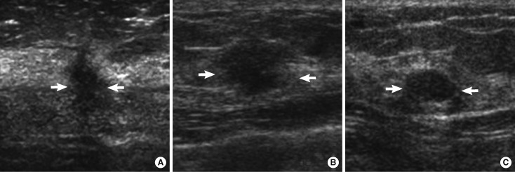
Purpose: To analyze significant ultrasonographic findings of small malignant breast mass (≤10 mm) which were occult on mammography.
Methods: The study included 190 small breast masses (≤10 mm), demonstrated on breast ultrasonography, but not mammography. Histopathology (when the masses were biopsied) or serial breast ultrasonography (for at least 24 months) were used to confirm benign or malignant condition of the masses. Univariate and multivariate logistic regression analysis were used to identify significant characteristic malignant findings on ultrasonography.
Results: Of 190 masses, 46 were cancer, and 144 were benign. On multivariate analyses, irregular shape (odds ratio [OR], 10.4) and not circumscribed margin (OR, 31.6) were significant features to differentiate between benign and malignant breast masses. However, low width/anteroposterior ratio, echogenic halo, hypoechogenecity and posterior acoustic shadow, which were predictors for malignancy in large breast mass, were not documented in small mass.
Conclusion: In conclusion, irregular shape and not circumscribed margin detected during ultrasonography were strong predictive signs of malignancy for small malignant breast mass.
Keywords: Breast; Carcinoma; Ultrasonography.
Conflict of interest statement
The authors declare that they have no competing interests.
Figures
References
-
- Smith RA, Cokkinides V, Brooks D, Saslow D, Brawley OW. Cancer screening in the United States, 2010: a review of current American Cancer Society guidelines and issues in cancer screening. CA Cancer J Clin. 2010;60:99–119. - PubMed
-
- Chao TC, Chen MF, Wang CS, Jan YY, Hwang TL, Chen SC. Small invasive breast carcinomas in Taiwanese women. Ann Surg Oncol. 2003;10:740–747. - PubMed
-
- Crystal P, Strano SD, Shcharynski S, Koretz MJ. Using sonography to screen women with mammographically dense breasts. AJR Am J Roentgenol. 2003;181:177–182. - PubMed
LinkOut - more resources
Full Text Sources