. 2012;7(10):e47357.
doi: 10.1371/journal.pone.0047357. Epub 2012 Oct 16.

Chloroquine or chloroquine-PI3K/Akt pathway inhibitor combinations strongly promote γ-irradiation-induced cell death in primary stem-like glioma cells

Affiliations

Chloroquine or chloroquine-PI3K/Akt pathway inhibitor combinations strongly promote γ-irradiation-induced cell death in primary stem-like glioma cells

Elke Firat et al. PLoS One. 2012.

Abstract

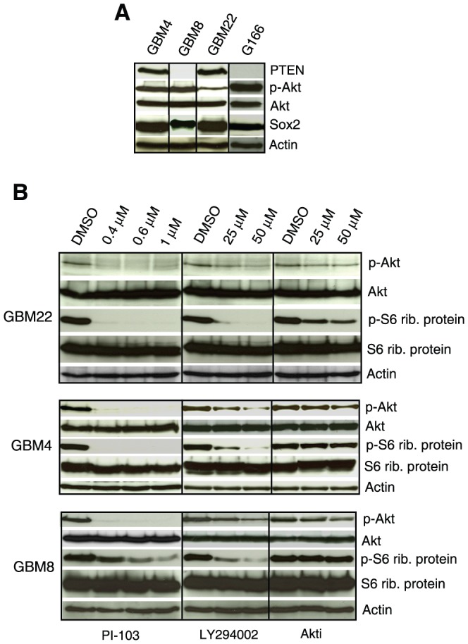
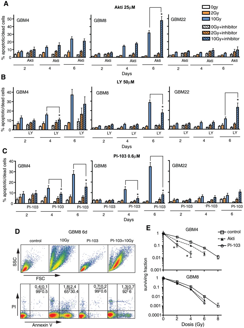
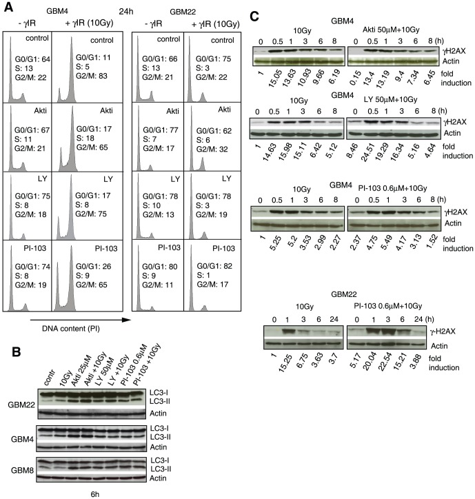
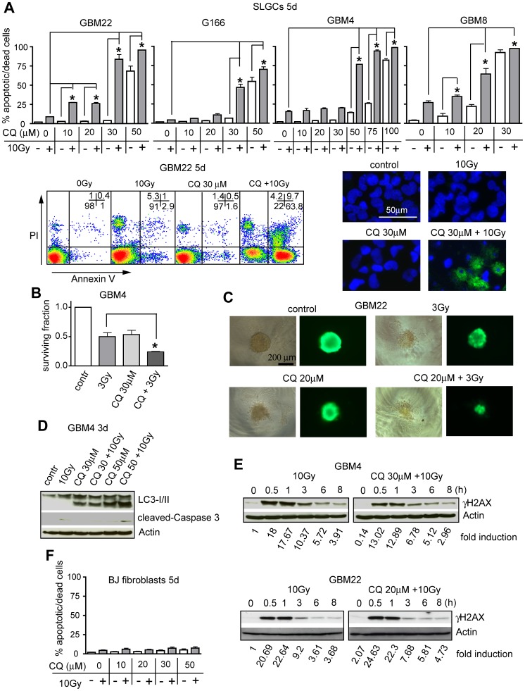
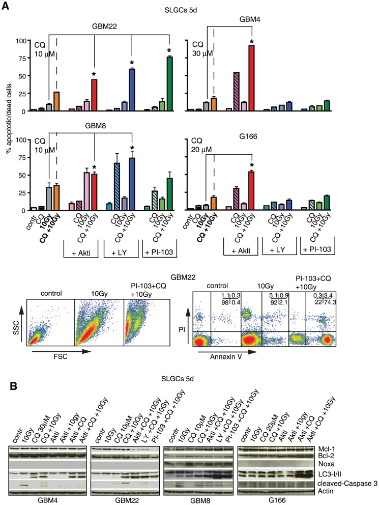
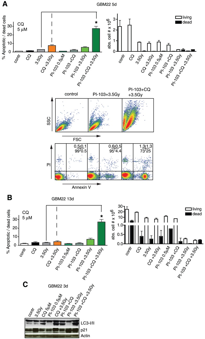
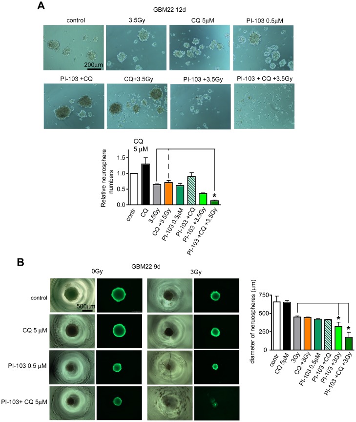
We asked whether inhibitors of the phosphatidylinositol 3-kinase (PI3K)/Akt pathway, which is highly active in cancer stem cells (CSCs) and upregulated in response to genotoxic treatments, promote γ-irradiationγIR)-induced cell death in highly radioresistant, patient-derived stem-like glioma cells (SLGCs). Surprisingly, in most cases the inhibitors did not promote γIR-induced cell death. In contrast, the strongly cytostatic Ly294002 and PI-103 even tended to reduce it. Since autophagy was induced we examined whether addition of the clinically applicable autophagy inhibitor chloroquine (CQ) would trigger cell death in SLGCs. Triple therapy with CQ at doses as low as 5 to 10 µM indeed caused strong apoptosis. At slightly higher doses, CQ alone strongly promoted γIR-induced apoptosis in all SLGC lines examined. The strong apoptosis in combinations with CQ was invariably associated with strong accumulation of the autophagosomal marker LC3-II, indicating inhibition of late autophagy. Thus, autophagy-promoting effects of PI3K/Akt pathway inhibitors apparently hinder cell death induction in γ-irradiated SLGCs. However, as we show here for the first time, the late autophagy inhibitor CQ strongly promotes γIR-induced cell death in highly radioresistant CSCs, and triple combinations of CQ, γIR and a PI3K/Akt pathway inhibitor permit reduction of the CQ dose required to trigger cell death.


Conflict of interest statement

Competing Interests: The authors have declared that no competing interests exist.

Figures

Figure 1
Figure 1. PI3K/Akt pathway activation status and inhibition of Akt phosphorylation by PI3K/Akt pathway inhibitors in primary SLGCs.
(A) Western blot analysis of basal Akt, p-Akt, and PTEN expression levels. Sox2 is shown as a stemness marker and β-Actin as a loading control. (B) Inhibition of phosphorylation of Akt (serine 473) and of S6 ribosomal protein by Akt inhibitor III (Akti), LY294002 and PI-103. The cells were treated with the inhibitors for 2 h before collecting the samples for analysis.
Figure 2
Figure 2. PI3K/Akt pathway inhibitors do not generally promote γIR-induced cell death in primary SLGCs.
(A–C) Percentage of annexin V/PI positive cells 2, 4, or 6 d after irradiation with 0, 2, or 10 Gy. Prior to irradiation, the cultures were pretreated for 1 h with Akt inhibitor III, LY294002 or PI-103. (D) Example of flow cytometric cell death analyses. The population with decreased forward light scatter (FSC) in the 10 Gy-irradiated sample reflects cell shrinkage and fragmentation typical of apoptosis. Early and mid apoptotic cells are annexin V-positive but still exclude PI; late apoptotic cells with compromised membrane integrity are annexin V/PI-double positive. (E) Clonogenic survival 14 d after treatment with γIR +/− Akt inhibitor III (25 µM) or PI-103 (0.6 µM). Experiments were performed in triplicates. Data in A–C represent means ± SD of three independent experiments. Statistical significance is indicated by an asterisk (p<0.05).
Figure 3
Figure 3. PI3K/Akt pathway inhibitors reduce G2M arrest and induce autophagy in γ-irradiated SLGCs.
(A) Cells were treated with Akt inhibitor III (25 µM), LY294002 (50 µM), or PI-103 (0.6 µM) for 1 h and then irradiated with 10 Gy. The cell cycle analyses were performed 24 h later. (B) Conversion of cytosolic LC3-I to autophagosome-associated LC3-II, and (C) kinetics of H2AX-phosphorylation, a measure of DNA damage; the SLGCs were pretreated with PI3K/Akt pathway inhibitors for 1 h before irradiation with 10 Gy. Representative Western blots are shown.
Figure 4
Figure 4. CQ and γIR synergistically induce strong apoptosis in SLGCs.
(A) Upper panel: percentage of annexin V/PI positive cells 5 d after treatment with CQ alone (white bars) or CQ plus γIR (gray bars). The cells were pretreated with CQ for 1 h and then irradiated with 10 Gy. Lower left: example of the flow cytometric analysis of apoptotic and dead cells; lower right: apoptotic morphology observed after Hoechst 33342/annexin V staining at 3 d after combination treatment. (B) Reduced survival of clonogenic cells 13 d after combination treatment. (C) Size reduction of preformed neurospheres 7 d after combination treatment. Viability of neurospheres was determined after calcein-AM staining. (D, E) Representative Western blots showing the effect of CQ or CQ plus γIR on expression levels of activated caspase-3 and LC3-I/II conversion (D), as well as on phosphorylation of H2AX (γH2AX) (E). (F) Lack of cell death induction in human fibroblasts. In A and F, data represent means of three independent experiments. Experiments in B and C were performed twice in triplicates. Statistical significance is indicated by an asterisk (p<0.05).
Figure 5
Figure 5. Triple combinations of γIR, a PI3K/Akt pathway inhibitor and low doses of CQ show additive to highly synergistic proapoptotic effects in primary SLGCs.
(A) Percentage of annexin V/PI positive cells 5 d after treatment with 10 Gy, a PI3K/Akt pathway inhibitor (25 µM Akt inhibitor III, 50 µM LY294002, and 0.6 µM PI-103) and CQ (at the doses indicated), alone, or in double or triple combinations. Lower panel: example of flow cytometric analysis of GBM22 SLGCs showing that most cells treated with the triple combinations die by apoptosis. (B) Western blot analyses showing a strong accumulation of LC3-II in SLGC samples with high proportions of annexin V/PI positive cells. In addition, expression levels of pro- and antiapoptotic molecules and cleaved caspase-3 are shown. Data in (A) are means of three independent experiments. Statistical significance is indicated by an asterisk (p<0.05).
Figure 6
Figure 6. Low doses of γIR, CQ and PI-103 synergistically induce apoptosis in GBM22 SLGCs.
(A, B) Left: Percentage of annexin V/PI positive cells 5 or 13 d after treatment with either γIR (3.5 Gy), CQ (5 µM) or PI-103 (0.5 µM) alone, with double or triple combinations. Right: cell numbers after trypan blue staining. Lower panel in A: flow cytometry data showing that cell death occurred primarily through apoptosis. (C) Western blot analysis of LC3-I/II conversion and of p21 expression levels.
Figure 7
Figure 7. Synergistic effects of a low dose triple combination including PI-103 in stem cell surrogate assays.
(A) Adherent monolayer cultures of GBM22 SLGCs were treated for 5 d as described in Fig. 6, then plated in serum-free medium to form neurospheres. Seven days later total neurospheres were counted and photographed. (B) Treatment of preformed 3D neurospheres. Cells were plated to form one neurosphere per well and 2 d later treated as indicated. 7 d after treatment, neurospheres were stained with calcein-AM to determine the viability and diameter of the spheres. Data in (A and B) are means of three independent experiments. Statistical significance is indicated by an asterisk (p<0.05).

References

    1. Stupp R, Mason WP, van den Bent MJ, Weller M, Fisher B, et al. (2005) Radiotherapy plus concomitant and adjuvant temozolomide for glioblastoma. N Engl J Med 352: 987–996. - PubMed
    1. Lathia JD, Venere M, Rao MS, Rich JN (2011) Seeing is believing: are cancer stem cells the Loch Ness monster of tumor biology? Stem Cell Rev 7: 227–237. - PMC - PubMed
    1. Maugeri-Sacca M, Vigneri P, De Maria R (2011) Cancer Stem Cells and Chemosensitivity. Clin Cancer Res 17: 4942–4947. - PubMed
    1. Fan QW, Weiss WA (2010) Targeting the RTK-PI3K-mTOR axis in malignant glioma: overcoming resistance. Curr Top Microbiol Immunol 347: 279–296. - PMC - PubMed
    1. Perry J, Okamoto M, Guiou M, Shirai K, Errett A, et al. (2012) Novel therapies in glioblastoma. Neurology Research International 2012: 428565. - PMC - PubMed

Publication types

MeSH terms